. 2011 Aug 31;31(35):12543-53.
doi: 10.1523/JNEUROSCI.1589-11.2011.

Accelerated age-related cognitive decline and neurodegeneration, caused by deficient DNA repair

Affiliations

Accelerated age-related cognitive decline and neurodegeneration, caused by deficient DNA repair

Nils Z Borgesius et al. J Neurosci. .

Abstract

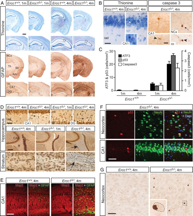
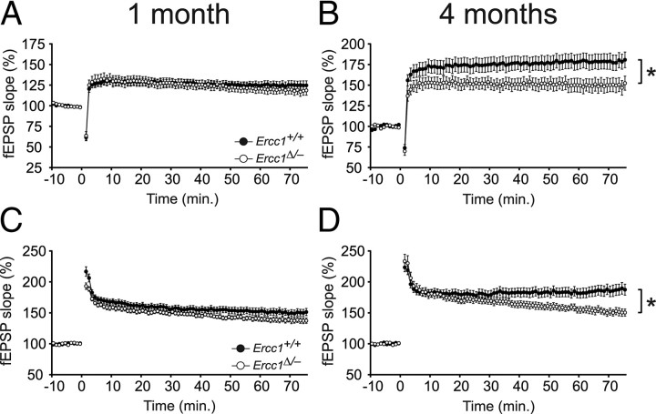
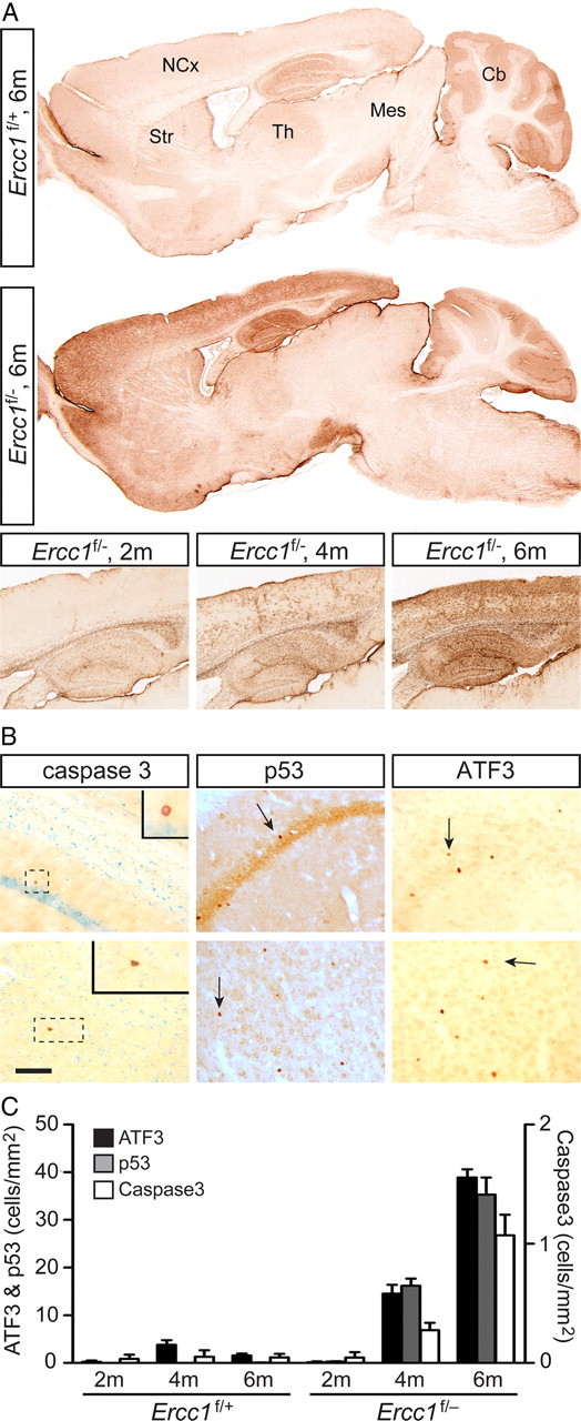
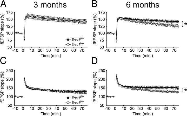
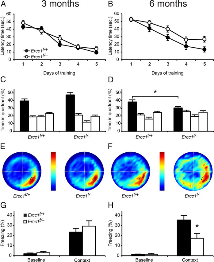
Age-related cognitive decline and neurodegenerative diseases are a growing challenge for our societies with their aging populations. Accumulation of DNA damage has been proposed to contribute to these impairments, but direct proof that DNA damage results in impaired neuronal plasticity and memory is lacking. Here we take advantage of Ercc1(Δ/-) mutant mice, which are impaired in DNA nucleotide excision repair, interstrand crosslink repair, and double-strand break repair. We show that these mice exhibit an age-dependent decrease in neuronal plasticity and progressive neuronal pathology, suggestive of neurodegenerative processes. A similar phenotype is observed in mice where the mutation is restricted to excitatory forebrain neurons. Moreover, these neuron-specific mutants develop a learning impairment. Together, these results suggest a causal relationship between unrepaired, accumulating DNA damage, and age-dependent cognitive decline and neurodegeneration. Hence, accumulated DNA damage could therefore be an important factor in the onset and progression of age-related cognitive decline and neurodegenerative diseases.


Figures

Figure 1.
Figure 1.
Young adult Ercc1Δ/− mice display reactive astrocytosis, mild neuronal degeneration, and signs of genotoxic stress. A, Coronal brain slices stained with thionine (upper panels) or processed for GFAP immunoperoxidase histochemistry (lower panels) illustrating normal gross histoarchitecture of the dorsal hippocampus and surrounding brain structures in Ercc1Δ/− mice, and increased GFAP staining throughout the brain of 4-month-old Ercc1Δ/− mice (scale bar, 500 μm). B, Light photomicrographs illustrating dying cells in hippocampus (CA1), cortex (NCx), and corpus callosum of 4-month-old Ercc1Δ/− mice. Dying cells are identified by their pyknotic nucleus in thionine-stained sections (white arrow and inset in second image), or by active caspase 3 staining. In many occasions, caspase 3-positive cells can be easily differentiated in neurons (arrows) or glial cells (arrowheads; scale bar, 25 μm). C, Quantification of cortical cell density positive for ATF3, p53 or caspase 3 (y-axis on left indicates values for ATF3 and p53; y-axis on the right indicates values for caspase 3). All data are reported as mean ± SEM. Two-way ANOVA revealed a significant effect for genotype, age and their interaction for ATF3 (all p < 0.0001), p53 (all p < 0.0001), and caspase 3 (all p < 0.01). D, Silver staining showing argyrophilic somatodendritic neuronal profiles indicative of dying neurons in 4-month-old Ercc1Δ/− hippocampus (arrows in upper row; scale bar, 50 μm), and argyrophilic degenerating axons in fimbria-fornix (ff) (arrow in middle row; scale bar, 250 μm) and striatal capsula interna (ci) bundles (arrow in lower row; scale bar, 50 μm) of 4-month-old Ercc1Δ/− brain. Note the absent and highly sporadic argyrophilic fiber degeneration in Ercc1+/+ mice and 1-month-old Ercc1Δ/− brain, respectively. E, Confocal immunofluorescent images of CA1 area in the hippocampus showing unaltered level of MAP2 and increased GFAP immunoreactivity in 4-month-old Ercc1Δ/− mice (scale bar, 100 μm). F, Confocal image showing p53-NeuN double-labeled cells in 4-month-old Ercc1Δ/− cortex and hippocampus (scale bar, 50 μm). G, ATF3-immunoperoxidase histochemistry illustrating multiple ATF3-positive cells in 4-month-old Ercc1Δ/− cortex. Inset shows enlargement of neuron with flattened eccentric nucleus (scale bar, 100 μm). Th, Thalamus; Str, striatum; Am, amygdala; cc, corpus callosum; DG, dentate gyrus; py, pyramidal layer; rad, stratum radiatum; ml, molecular layer; gr, granule layer.
Figure 2.
Figure 2.
Ercc1Δ/− mice show reduced synaptic plasticity at 4 months of age. A, LTP (10 Hz) in 1-month-old mice shows no difference between Ercc1Δ/− and Ercc1+/+ mice (n = 14 slices from 9 animals and n = 12 slices from 8 animals for Ercc1+/+ and Ercc1Δ/−, respectively). B, LTP (10 Hz) in 4-month-old mice shows reduced LTP in Ercc1Δ/− mice [n = 19 slices from 10 animals and n = 18 slices from 11 animals (4 males and 6 females) for Ercc1+/+ and Ercc1Δ/−, respectively]. C, LTP (100 Hz) in 1-month-old mice shows no difference between Ercc1Δ/− and Ercc1+/+ mice (n = 17 slices from 9 animals and n = 12 slices from 8 animals for Ercc1+/+ and Ercc1Δ/−, respectively). D, LTP (100 Hz) in 4-month-old mice shows reduced LTP in Ercc1Δ/− mice (n = 18 slices from 10 animals and n = 17 slices from 11 animals and for Ercc1+/+ and Ercc1Δ/−, respectively). All data are reported as mean ± SEM. Filled circles represent Ercc1+/+. Open circles represent Ercc1Δ/−. *Significantly different (p < 0.05) from age-matched Ercc1+/+ mice.
Figure 3.
Figure 3.
Ercc1f/− mice show astrocytosis, genotoxic stress transcription factor expression, and mild age-related neuronal degeneration. A, GFAP-immunoperoxidase staining in sagittal brain section of Ercc1f/− and control (Ercc1f/+) mice. Note the strongly increased GFAP staining in forebrain regions, including neocortex (NCx), striatum (Str), and hippocampus, of 6-month-old Ercc1f/− mice while GFAP staining in other parts of the brain, including thalamus (Th), mesencephalon (Mes), and cerebellum (Cb) is the same as in control. The lower panels show that the increase GFAP staining in Ercc1f/− hippocampus and cortex starts after 2 months of age and is progressive. B, Active caspase 3, p53, and ATF3-immunoperoxidase histochemistry illustrating positive cells in 6-month-old Ercc1f/− hippocampal CA1 area (upper row) and neocortex (lower row, scale bar 100 μm). C, Bar graph illustrating the density of ATF3- and p53- (y-axis on left) and caspase 3- (y-axis on the right) positive cells in neocortex of Ercc1f/− and Ercc1f/+ mice. Data represent means ± SEM. Two-way ANOVA revealed a significant effect for genotype, age, and their interaction for ATF3 (all p < 0.001), p53 (all p < 0.001), and caspase 3 (genotype p = 0.01, age p = 0.01 and genotype × age p = 0.01). Tukey HSD post hoc tests revealed significant effects between all age groups for ATF3 (all p < 0.001) and for p53 (all p < 0.001). For caspase 3, Tukey HSD post hoc test revealed a significant difference between 2 and 6 months and 4 and 6 months (p < 0.001 and p = 0.002, respectively).
Figure 4.
Figure 4.
Ercc1f/− mice show reduced synaptic plasticity at 6 months of age. A, LTP (10 Hz) in 3-month-old mice shows no difference between Ercc1f/− and Ercc1f/+ mice [n = 16 slices from 5 animals, n = 19 slices from 8 animals for Ercc1f/+ and Ercc1f/−, respectively]. B, LTP (10 Hz) in 6-month-old mice shows reduced LTP in Ercc1f/− mice (n = 14 slices from 4 animals and n = 11 slices from 5 animals for Ercc1f/+ and Ercc1f/−, respectively). C, LTP (100 Hz) in 3-month-old mice shows no difference between Ercc1f/− and Ercc1f/+ mice (n = 13 slices from 4 animals, n = 18 slices from 10 animals for Ercc1f/+ and Ercc1f/−, respectively). D, LTP (100 Hz) in 6-month-old mice shows reduced LTP in Ercc1f/− mice (n = 15 slices from 5 animals and n = 17 slices from 7 animals for Ercc1f/+ and Ercc1f/−, respectively). All data are reported as mean ± SEM. Filled circles represent Ercc1f/+. Open circles represent Ercc1f/−. *Significantly different (p < 0.05) from age-matched Ercc1f/+ mice.
Figure 5.
Figure 5.
Ercc1f/− mice show impaired fear conditioning and impaired water maze performance at 6 months of age. A, B, Escape latency of 3-month-old (A) and 6-month-old (B) mice shows no difference between Ercc1f/− and Ercc1f/+ mice (n = 9, n = 15, n = 11, and n = 17 for 3- and 6-month-old Ercc1f/+ and Ercc1f/−, respectively). Filled circles represent Ercc1f/+. Open circles represent Ercc1f/−. C, D, Quantification of quadrant occupancy in 3-month-old (C) and 6-month-old (D) mice, during the probe trial performed at day 5. Although all groups searched significantly more in the target quadrant, at 6 months old, Ercc1f/− mice spent significantly less time in the target quadrant than their Ercc1f/+ littermates. Black bar represents target quadrant. E, F, Visual representation of all search tracks during the probe trial of the 3-month-old (E) and 6-month-old (F) mice. The color of the heat plots indicate the mean time spent at a certain location. The white and black dashed lines indicate quadrants and former platform location, respectively. G, H, Contextual fear conditioning in 3-month-old mice (n = 9 and n = 20 for Ercc1f/+ and Ercc1f/−, respectively) (G) and 6-month-old mice (n = 10 and n = 13 for Ercc1f/+ and Ercc1f/−, respectively) (H). Six-month-old Ercc1f/− mice show normal baseline freezing before the shock but significantly reduced freezing when placed in the context 24 h after the shock. All data are reported as mean ± SEM. *Significantly different (p < 0.05) from age-matched Ercc1f/+ mice.

Similar articles

Cited by

References

    1. Ahles TA, Saykin AJ. Candidate mechanisms for chemotherapy-induced cognitive changes. Nat Rev Cancer. 2007;7:192–201. - PMC - PubMed
    1. Ahmad A, Robinson AR, Duensing A, van Drunen E, Beverloo HB, Weisberg DB, Hasty P, Hoeijmakers JH, Niedernhofer LJ. ERCC1-XPF endonuclease facilitates DNA double-strand break repair. Mol Cell Biol. 2008;28:5082–5092. - PMC - PubMed
    1. Bach ME, Barad M, Son H, Zhuo M, Lu YF, Shih R, Mansuy I, Hawkins RD, Kandel ER. Age-related defects in spatial memory are correlated with defects in the late phase of hippocampal long-term potentiation in vitro and are attenuated by drugs that enhance the cAMP signaling pathway. Proc Natl Acad Sci U S A. 1999;96:5280–5285. - PMC - PubMed
    1. Barnes CA. Long-term potentiation and the ageing brain. Philos Trans R Soc Lond B Biol Sci. 2003;358:765–772. - PMC - PubMed
    1. Bergstralh DT, Sekelsky J. Interstrand crosslink repair: can XPF-ERCC1 be let off the hook? Trends Genet. 2008;24:70–76. - PubMed

Publication types

MeSH terms