. 2011 Aug 31;31(35):12554-65.
doi: 10.1523/JNEUROSCI.3143-11.2011.

Kalirin binds the NR2B subunit of the NMDA receptor, altering its synaptic localization and function

Affiliations

Kalirin binds the NR2B subunit of the NMDA receptor, altering its synaptic localization and function

Drew D Kiraly et al. J Neurosci. .

Abstract

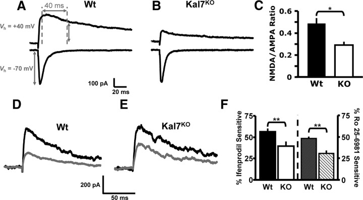
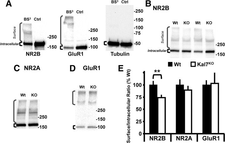
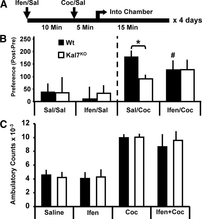
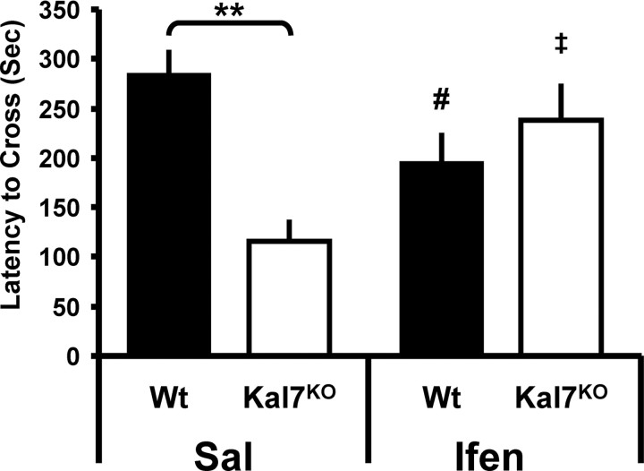
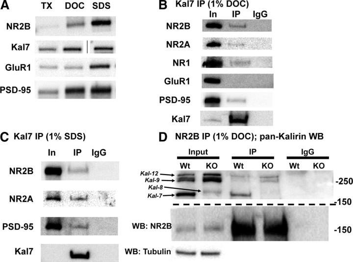
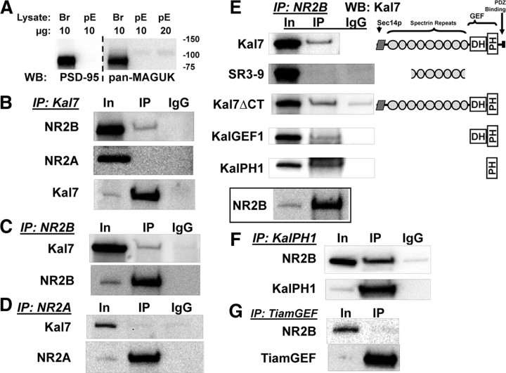
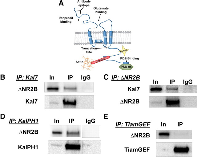
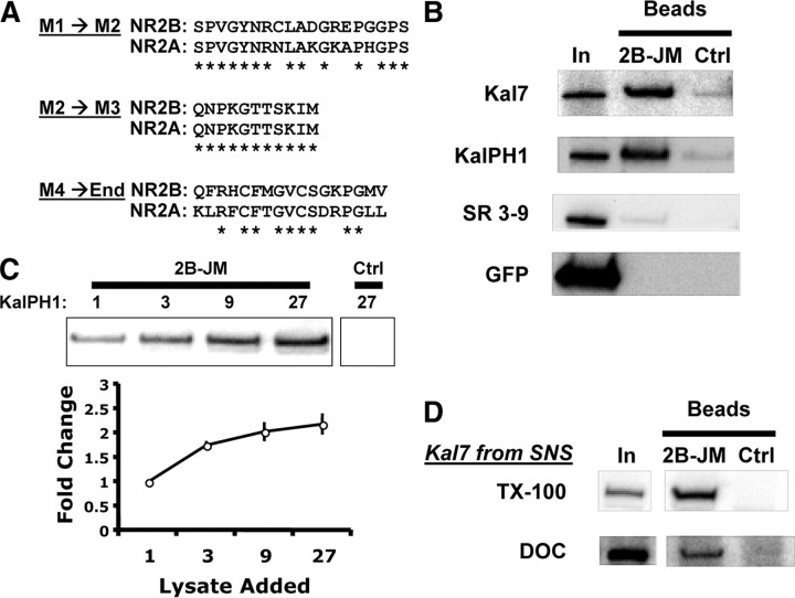
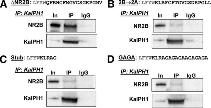
The ability of dendritic spines to change size and shape rapidly is critical in modulating synaptic strength; these morphological changes are dependent upon rearrangements of the actin cytoskeleton. Kalirin-7 (Kal7), a Rho guanine nucleotide exchange factor localized to the postsynaptic density (PSD), modulates dendritic spine morphology in vitro and in vivo. Kal7 activates Rac and interacts with several PSD proteins, including PSD-95, DISC-1, AF-6, and Arf6. Mice genetically lacking Kal7 (Kal7(KO)) exhibit deficient hippocampal long-term potentiation (LTP) as well as behavioral abnormalities in models of addiction and learning. Purified PSDs from Kal7(KO) mice contain diminished levels of NR2B, an NMDA receptor subunit that plays a critical role in LTP induction. Here we demonstrate that Kal7(KO) animals have decreased levels of NR2B-dependent NMDA receptor currents in cortical pyramidal neurons as well as a specific deficit in cell surface expression of NR2B. Additionally, we demonstrate that the genotypic differences in conditioned place preference and passive avoidance learning seen in Kal7(KO) mice are abrogated when animals are treated with an NR2B-specific antagonist during conditioning. Finally, we identify a stable interaction between the pleckstrin homology domain of Kal7 and the juxtamembrane region of NR2B preceding its cytosolic C-terminal domain. Binding of NR2B to a protein that modulates the actin cytoskeleton is important, as NMDA receptors require actin integrity for synaptic localization and function. These studies demonstrate a novel and functionally important interaction between the NR2B subunit of the NMDA receptor and Kalirin, proteins known to be essential for normal synaptic plasticity.


Figures

Figure 1.
Figure 1.
NMDA/AMPA receptor current ratio and NR2B subunit-containing NMDA receptor-mediated currents are diminished in Kal7KO neurons. AC. To measure NMDA/AMPA receptor current ratio in layer 2/3 pyramidal neurons, the AMPA receptor response was quantified as the peak current at a holding potential of −70 mV; the NMDA receptor response was quantified using a holding potential of +40 mV and was measured 40 ms after stimulation onset. A, B, Representative evoked EPSCs from Wt and Kal7KO mice at these holding potentials. C, Group data for the NMDA/AMPA receptor current ratio in WT and Kal7KO mice (*p < 0.05; t test, n = 8 mice per genotype). D–F, To measure NR2B subunit-containing NMDA receptor-mediated currents, layer 2/3 pyramidal neurons were voltage clamped at +50 mV in the presence of 10 μm DNQX to block AMPA receptor-mediated currents. D, E, Representative evoked current traces from both genotypes are shown; black traces were made at baseline and gray traces were made 15 min after bath application of an NR2B subunit-specific antagonist, ifenprodil (3 μm). F, Mean data demonstrate that Wt animals have a significantly larger portion of their NMDA receptor current that is sensitive to ifenprodil (left) or Ro 25-6981 (right; 0.5 μm) a different NR2B subunit-specific antagonist. (ifenprodil, **p < 0.01; t test, n = 5 animals/group, 2–3 cells/animal; Ro 25–6981, **p < 0.01; t test, n = 3–4 animals/group, 2–3 cells/animal).
Figure 2.
Figure 2.
Knock-out of Kal7 specifically decreases surface localization of NR2B subunit. A, Exposure of cortical slices kept at 4°C to BS3 cross-links surface receptors, producing high molecular weight bands that are not observed in non-cross-linked control (Ctrl) slices; brackets indicate Surface and Intracellular receptors. As expected, an intracellular protein, tubulin, was not cross-linked. B–D, Representative Western blots of cross-linked samples from Wt and Kal7KO slices blotted for NR2B (B), NR2A (C), and GluR1 (D). E, Surface and Intracellular receptor levels were quantified for each sample, and the Surface/Intracellular ratio for Wt slices was normalized to 100%. Kal7KO animals show a decrease in cell surface expression of NR2B, while surface levels of NMDA receptor subunit NR2A and the AMPA receptor subunit GluR1 were unchanged. (**p = 0.018 as calculated by t test; N = 9–10 slices/genotype, 3 animals/group).
Figure 3.
Figure 3.
Blockade of NR2B subunit-containing NMDA receptors eliminates differences in cocaine-conditioned place preference response in Kal7KO and Wt animals. A, Timeline of daily injections. Cocaine (Coc) injections (10 mg/kg) were preceded by ifenprodil (Ifen) injections (2 mg/kg, i.p.) to block NR2B subunit-containing NMDA receptors. Sal, Saline. B, Left, Animals that received saline only or ifenprodil before saline showed no genotypic differences and no significant preference/aversion (n = 3–5/group). B, Right, In the groups receiving saline injections before cocaine injections, Kal7KO animals showed a robust decrease in place preference (effect of genotype within saline, *p = 0.02; Holm–Sidak test). In Wt animals, when ifenprodil preceded the cocaine injection there was a significant decrease in preference compared to saline pretreatment (effect of treatment within Wt, #p = 0.01). The genotypic difference between Wt and Kal7KO animals was abrogated when animals were pretreated with ifenprodil (effect of genotype within ifenprodil, p = 0.61) (n = 7–10/group). C, Acute locomotor effects of cocaine (10 mg/kg), ifenprodil (2 mg/kg), or both administered concomitantly. Ifenprodil alone produced no effect on locomotor activity. While cocaine produced the expected increase in locomotor activity, simultaneous injection of ifenprodil did not alter the response. There were no genotype-dependent effects in this study (n = 4/group).
Figure 4.
Figure 4.
Blockade of NR2B subunit-containing NMDA receptors abrogates genotypic differences in passive avoidance fear conditioning. Left, In animals receiving an injection of saline (Sal) before passive avoidance conditioning, Kal7KO animals showed significantly decreased conditioning compared to Wt mice (**p = 0.003; Holm–Sidak test). Right, In animals treated with ifenprodil (Ifen) before conditioning, the genotypic difference was eliminated (p = 0.18). While Wt animals showed a significant decrease in conditioning with ifenprodil pretreatment (effect of treatment within Wt, #p = 0.01), Kal7KO animals exhibited a significant increase in conditioning following NR2B blockade (effect of treatment within Wt, p = 0.04) (n = 6–12/group).
Figure 5.
Figure 5.
Kal7 associates with NMDA receptor complexes in vivo. A, Solubilization of rat forebrain synaptosomal proteins with different detergents yielded different amounts of soluble NR2B, Kal7, GluR1, and PSD-95. Samples were loaded as equal percentages of the supernatant after detergent extraction; line in Kal7 sample indicates image is from a nonadjacent well of the same gel. B, In samples solubilized with DOC, immunoprecipitation (IP) with the Kal7 mAb coprecipitates a small fraction of the NR2B, NR2A, NR1, and PSD-95, but no detectable GluR1. Incubation of sample with preimmune IgG showed no binding. IP came from 50-fold more sample than Input (In); recovery of Kal7 is shown in the bottom panel. C, Solubilization of synaptosomes with 1% SDS revealed coprecipitation of NMDAR subunits and PSD-95 with Kal7; IgG controls again showed no binding. IP came from 100-fold more sample than Input; recovery of Kal7 is shown in the bottom panel. D, Synaptosomes from Wt and Kal7KO animals were solubilized with DOC, immunoprecipitated using an NR2B antibody, and blotted for all isoforms of Kalirin. Examination of the Input sample confirmed the absence of Kal7 and increase in Kal8/9/12 in Kal7KO animals, as reported (Ma et al., 2008b). A fraction of the Kal7 coimmunoprecipitated with NR2B in Wt extracts, as did small amounts of Kal9 and Kal12. In Kal7KO samples, coimmunoprecipitation of Kal9 and Kal12 was more apparent. Western blotting (WB) for tubulin confirmed equal loading in the input lanes, and IgG controls confirmed the absence of nonspecific binding; recovery of NR2B is shown in the middle panel. Similar results were observed in triplicate and quadruplicate (B, C) or duplicate (D) samples.
Figure 6.
Figure 6.
KalPH1 interacts with NR2B. A, pEAK Rapid cell (pE) and mouse brain (Br) lysate (10 or 20 μg of protein) were blotted for PSD-95 and other MAGUK scaffolding proteins. B, In pEAK Rapid cells cotransfected with vectors encoding NR1, NR2B, and Kal7 (top) or NR1, NR2A, and Kal7 (middle), immunoprecipitation (IP) with Kal7 mAb revealed bound NR2B but no bound NR2A. Samples in B–D were loaded as Input (IN)(1):IP(90). In cells cotransfected with NR1, NR2B, and Kal7 (C), immunoprecipitation with antibody to the extracellular domain of NR2B coprecipitated a small percentage of the Kal7; recovery of NR2B is shown in the bottom panel. In cells transfected with NR1, NR2A, and Kal7 (D), antibody to NR2A did not coprecipitate Kal7; recovery of NR2A is shown in the bottom panel. IgG controls showed no nonspecific binding. E, To map the interaction sites in Kal7, cells were cotransfected with vectors encoding NR1, NR2B, and the indicated fragments of Kal7. Cells were extracted in DOC and NR2B was immunoprecipitated; Kalirin fragments were then probed for coprecipitation using the appropriate antibody. The KalPH1 domain is sufficient for Kal7 coprecipitation with NR2B. Recovery of NR2B is shown in the bottom panel and IgG controls are indicated. WB, Western blotting. F, pEAK Rapid cells were cotransfected with vectors encoding NR1, NR2B, and KalPH1-GFP. GFP immunoprecipitates were probed for NR2B. G, Interaction specificity was assessed by cotransfecting vectors encoding NR1, NR2B, and HA-tagged GEF domain of Tiam-1 (TiamGEF), another Rac-GEF localized to the PSD. IP of TiamGEF (using HA antibody) did not coprecipitate NR2B. Samples for F and G were loaded as Input(1):IP(90), with recoveries shown in the bottom panels. IgG controls showed no background staining; each of these IPs was performed on 3–7 sample preparations with similar results each time.
Figure 7.
Figure 7.
Interaction between KalPH1 and NR2B involves a membrane proximal intracellular segment of NR2B. A, Diagram of the NR2B subunit showing the C terminus of ΔNR2B (residues 1–861 of NR2B) (Foster et al., 2010) and the antibody binding epitope. Sites known to interact with specific proteins are indicated. B, pEAK Rapid cells cotransfected with vectors encoding Kal7 and ΔNR2B were immunoprecipitated with Kal7 mAb beads; coprecipitated ΔNR2B was visualized using monoclonal antibody to the extracellular domain. All samples shown in this figure were cotransfected with NR1 and loaded Input (In)(1):IP(90); recoveries are shown in the bottom panels. IP, Immunoprecipitation. C, Immunoprecipitation of ΔNR2B coprecipitated Kal7, which was visualized with Kal7 polyclonal antibody. D, Cells cotransfected with vectors encoding KalPH1-GFP and ΔNR2B were immunoprecipitated with rabbit polyclonal antibody to GFP; the coprecipitated ΔNR2B was visualized using monoclonal antibody to the extracellular domain, indicating that this interaction is specific to the KalPH1 domain. E, Cells cotransfected with vectors encoding HA-tagged TiamGEF and ΔNR2B were immunoprecipitated with polyclonal antibody to HA; TiamGEF does not coprecipitate ΔNR2B. IgG controls showed no background binding. All IPs were repeated at least four times with similar results.
Figure 8.
Figure 8.
Interaction of KalPH1 with the juxtamembrane region of NR2B A, Clustal analysis comparing intracellular regions of NR2B and NR2A; M1, M2, M3, and the C terminus of ΔNR2B (End) are as shown in Fig. 7A. B, Lysates from cells expressing the indicated proteins were incubated with NR2B-JM or control (no linked peptide) beads. The 2B-JM beads bound Kal7 and KalPH1-GFP but did not bind SR3-9 or GFP. None of the constructs bound to control beads. In, Input; Ctrl, control. C, Increasing amounts of KalPH1-GFP lysate were incubated with a fixed volume of 2B-JM or control beads; binding of KalPH1-GFP began to saturate as the amount of lysate was increased; error bars are standard deviation of triplicates. Control beads incubated with the highest amount of KalPH1-GFP lysate showed no evidence of binding. D, 2B-JM beads were incubated with mouse forebrain synaptosomes solubilized with 1% TX-100 or 1% DOC (dialyzed). Kal7 solubilized in either way bound to 2B-JM beads but not to control beads. Pulldowns for B and D were performed in duplicate or triplicate with similar results.
Figure 9.
Figure 9.
Mutation of the 2B-JM region decreases KalPH1-ΔNR2B coprecipitation. The sequences of the final juxtamembrane region of ΔNR2B (A), the NR2B→2A mutant (B), the Stub mutant (C) and the GAGA mutant (D) are shown (black text) with the preceding four amino acids (gray text). Cells cotransfected with NR1, each ΔNR2B construct, and KalPH1-GFP were immunoprecipitated with antibody to GFP or control IgG. Coprecipitation of ΔNR2B was observed as shown in previous figures; each mutation reduced coprecipitation to background levels. Images for A–C were all from the same gel; images for D were from a different gel exposed for the same time. ΔNR2B and all mutants equally coprecipitated NR1, indicating the formation of stable receptor complexes (data not shown). In, Input; IP, immunoprecipitation.

Similar articles

Cited by

References

    1. Akashi K, Kakizaki T, Kamiya H, Fukaya M, Yamasaki M, Abe M, Natsume R, Watanabe M, Sakimura K. NMDA receptor GluN2B (GluR epsilon 2/NR2B) subunit is crucial for channel function, postsynaptic macromolecular organization, and actin cytoskeleton at hippocampal CA3 synapses. J Neurosci. 2009;29:10869–10882. - PMC - PubMed
    1. Al-Hallaq RA, Conrads TP, Veenstra TD, Wenthold RJ. NMDA di-heteromeric receptor populations and associated proteins in rat hippocampus. J Neurosci. 2007;27:8334–8343. - PMC - PubMed
    1. Allison DW, Gelfand VI, Spector I, Craig AM. Role of actin in anchoring postsynaptic receptors in cultured hippocampal neurons: differential attachment of NMDA versus AMPA receptors. J Neurosci. 1998;18:2423–2436. - PMC - PubMed
    1. Barria A, Malinow R. NMDA receptor subunit composition controls synaptic plasticity by regulating binding to CaMKII. Neuron. 2005;48:289–301. - PubMed
    1. Benavides DR, Quinn JJ, Zhong P, Hawasli AH, DiLeone RJ, Kansy JW, Olausson P, Yan Z, Taylor JR, Bibb JA. Cdk5 modulates cocaine reward, motivation, and striatal neuron excitability. J Neurosci. 2007;27:12967–12976. - PMC - PubMed

Publication types

MeSH terms