. 2011 Dec;301(6):C1351-9.
doi: 10.1152/ajpcell.00166.2011. Epub 2011 Aug 31.

TM4SF10 and ADAP interaction in podocytes: role in Fyn activity and nephrin phosphorylation

Affiliations

TM4SF10 and ADAP interaction in podocytes: role in Fyn activity and nephrin phosphorylation

Timur A Azhibekov et al. Am J Physiol Cell Physiol. 2011 Dec.

Abstract

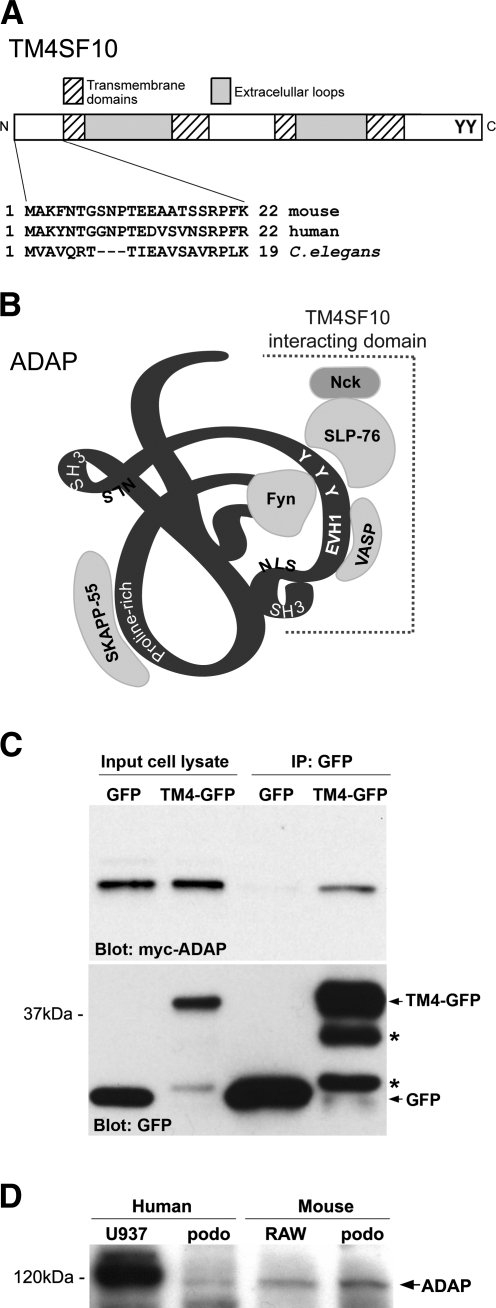
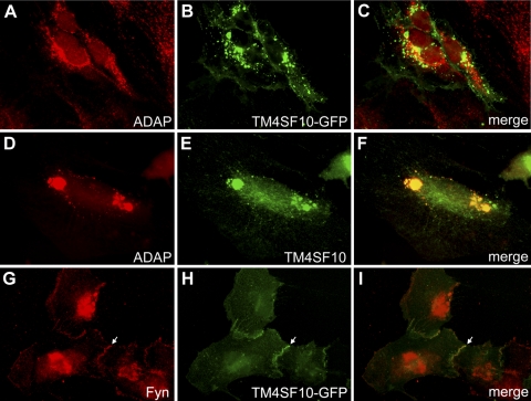
TM4SF10 [transmembrane tetra(4)-span family 10] is a claudin-like cell junction protein that is transiently expressed during podocyte development where its expression is downregulated in differentiating podocytes coincident with the appearance of nephrin at the slit diaphragm. In a yeast two-hybrid screen, we identified adhesion and degranulation-promoting adaptor protein (ADAP), a well-known Fyn substrate and Fyn binding partner, as a TM4SF10 interacting protein in mouse kidney. Using coimmunoprecipitation and immunohistochemistry experiments in cultured human podocytes, we show that TM4SF10 colocalizes with Fyn and ADAP but does not form a stable complex with Fyn. Cytoskeletal changes and phosphorylation events mediated by Fyn activity were reversed by TM4SF10 overexpression, including a decrease in the activating tyrosine phosphorylation of Fyn (Y(421)), suggesting TM4SF10 may have a regulatory role in suppressing Fyn activity. In addition, TM4SF10 was reexpressed following podocyte injury by puromycin aminonucleoside treatment, and its expression enhanced the abundance of high-molecular-weight forms of nephrin indicating it may participate in a mechanism controlling nephrin's appearance at the plasma membrane. Therefore, these studies have identified ADAP as another Fyn adapter protein expressed in podocytes, and that TM4SF10, possibly through ADAP, may regulate Fyn activity. Since TM4SF10 expression is temporally regulated during kidney development, these studies may help define a mechanism by which the slit diaphragm matures as a highly specialized cell junction during podocyte differentiation.


Figures

Fig. 1.
Fig. 1.
Adhesion and degranulation-promoting adaptor protein (ADAP) is a TM4SF10 [transmembrane tetra(4)-span family 10] interacting protein and is expressed in podocytes. A: schematic diagram of TM4SF10 protein domain structure noting the highly conserved 22 amino acid NH2-terminus (N) used as bait in the yeast two hybrid screen, the four trans-membrane domains typical of the tetraspanin proteins, and potential tyrosine phosphorylation sites on the COOH-terminus (C). B: schematic ribbon diagram of the 130-kDa ADAP protein (not drawn to scale) showing protein-protein interaction motifs (proline-rich, SH3, and EVH1), putative nuclear localization signal (NLS), and known tyrosine (Y) phosphorylation sites in addition to proteins that bind ADAP in lymphocytes (Fyn, VASP, SKAPP-55, and SLP-76 which also binds Nck). The region identified in the yeast two-hybrid screen and putative binding region for TM4SF10 is shown with the dashed line and span amino acids 378 to the COOH-terminus. C: Western blot of cell lysates from MDCK cells stably expressing GFP (lanes 1, 3) or TM4SF10-green fluorescent protein (GFP) (“TM4-GFP,” lanes 2, 4), and cotransfected with myc-ADAP. Cell lysates were immunoprecipitated with anti-GFP antibodies (lanes 3, 4) and blotted for myc (top) or GFP (bottom); *translationally truncated or proteolytically cleaved TM4SF10-GFP fusion proteins. D: Western blot of whole cell lysates from human and mouse podocyte (“podo”) cell lines confirming ADAP expression. Murine monocytic cell line RAW264.7 (RAW) and human monocytic cell line U937 were used as positive controls.
Fig. 2.
Fig. 2.
TM4SF10 colocalizes with ADAP and Fyn in podocytes. Detection by immunohistochemistry of TM4SF10-GFP (B,H) or endogenous TM4SF10 (E) in podocytes and colocalization with endogenous ADAP (A,D) or endogenous Fyn (G); respective merged images in C, F, and I. TM4SF10 was originally described as an endoplasmic reticulum (ER) and plasma membrane protein (4), and both endogenous and GFP-tagged TM4SF10 localized to intracellular compartments and the plasma membrane, with concentrations at cell-cell contacts (arrow).
Fig. 3.
Fig. 3.
TM4SF10 coimmunoprecipitates with ADAP but not with Fyn in podocytes. A: ADAP interactions with Fyn and TM4SF10 were confirmed in podocytes stably expressing TM4SF10-GFP (“TM4-GFP”) by immunoprecipitating with Fyn, ADAP, or GFP antibodies and Western blotted for ADAP (“input”, whole cell lysates). B: Fyn interactions with TM4SF10 were tested in similar immunoprecipitation experiments, using podocytes stably expressing TM4SF10-GFP (TM4-GFP) or GFP (as a negative control), after transient transfection with Fyn plasmid. Cell lysates were immunoprecipitated with either: a control species and isotype matched IgG (IgG); an anti-Fyn antibody (Fyn); or an anti-GFP antibody (GFP), and Western blotted for either Fyn or GFP. Some of the IgG heavy chain of the immunoprecipitating antibody is observed in these blots due to cross reactivity with the Western blotting antibody.
Fig. 4.
Fig. 4.
TM4SF10 functionally alters cell shape and Fyn activity in MDCK cells. A: MDCK cells stably expressing GFP, TM4SF10-GFP, or Fyn-GFP, and coexpression of TM4SF10-GFP in Fyn-GFP expressing cells were examined by phase-contrast microscopy to monitor changes in colony formation behavior. B: MDCK cells expressing the same GFP fusion proteins as shown in A were plated at low density to visual morphology of individual cells. Representative cell images are shown and percentage of cells with extensions are graphed below (*P < 0.001 compared with all other samples, **P < 0.001 compared with Fyn-GFP). C: MDCK cells stably expressing Fyn-GFP were cotransfected with TM4SF10-GFP or ADAP-GFP, followed by immunoblotting with an anti-phospho-tyrosine antibody (P-Tyr) to show total cellular tyrosine kinase activity. D: schematic diagram of Fyn kinase and two major phosphorylation sites: the activating phospho-tyrosine421 (pY421) of the kinase domain and the inactivating phospho-tyrosine532 (pY532) that induces conformation changes that block the SH2 domain. E: effect of TM4SF10-GFP on Fyn phosphorylation using Western blotting for total Fyn and phospho-specific antibodies. The additional band on the Src pY421 blot (which is not detected by the Fyn-specific antibody) is likely another Src family kinase since these antibodies will detect the same phospho-epitope on other Src kinases; however, the degree of phosphorylation of this other band does not appear to change with TM4SF10-GFP expression.
Fig. 5.
Fig. 5.
TM4SF10 colocalizes with nephrin and is associated with changes in nephrin abundance and phosphorylation in podocytes. AC: immunohistochemistry for F-actin (Phalloidin) comparing a podocyte with and without TM4SF10-GFP expression. Cells expressing TM4SF10-GFP cells had more circumferential bands (B, arrows) of F-actin, whereas cells without TM4SF10-GFP had a more typical stress fiber appearance of F-actin cross striations. DG: colocalization of TM4SF10-GFP and nephrin in podocytes. EG: higher magnification of boxed region in D showing partial colocalization of nephrin and TM4SF10-GFP in fine, distal cell extension. H: Western blot of podocyte nephrin expression following overexpression or knockdown of TM4SF10-GFP using a TM4SF10-specific short hairpin RNA (shRNA). The high-molecular-weight forms of nephrin at 175 and 185 kDa are noted with arrows. Efficiency of shRNA knockdown of TM4SF10 was confirmed by Western blot with anti-GFP antibodies and Tubulin was used as a loading control. I: Western blot of total nephrin abundance and presence of nephrin tyrosine1217 phosphorylation (pY1217) with and without TM4SF10-GFP overexpression and with and without preincubation with pervanadate to block tyrosine phosphatase activity.
Fig. 6.
Fig. 6.
Puromycin aminonucleoside (PAN) treatment induces reexpression of TM4SF10. A: cultured podocytes were treated with PAN or phosphate-buffered saline as a control and were immunostained for native expression of TM4SF10 (green) and cadherin (red), along with the nuclear stain DAPI (blue). Note the simplification of interdigitations of cell-cell contacts (cadherin-positive cell junctions) with PAN treatment compared with the control treated cells. B: immunohistochemistry for TM4SF10 (green) in kidney sections from rats treated with PAN or saline as a control using the standard PAN nephrosis model of podocyte injury. In this model, postinjection day 2 is a time point before the onset of severe proteinuria, whereas day 10 is a time point where rats exhibit nephrotic range proteinuria. Panels are matched exposures; ×40 magnification.

References

    1. Behrens J, Vakaet L, Friis R, Winterhager E, Van RF, Mareel MM, Birchmeier W. Loss of epithelial differentiation and gain of invasiveness correlates with tyrosine phosphorylation of the E-cadherin/beta-catenin complex in cells transformed with a temperature-sensitive v-SRC gene. J Cell Biol 120: 757–766, 1993 - PMC - PubMed
    1. Blasutig IM, New LA, Thanabalasuriar A, Dayarathna TK, Goudreault M, Quaggin SE, Li SS, Gruenheid S, Jones N, Pawson T. Phosphorylated YDXV motifs and Nck SH2/SH3 adaptors act cooperatively to induce actin reorganization. Mol Cell Biol 28: 2035–2046, 2008 - PMC - PubMed
    1. Bruggeman LA, Martinka S, Simske JS. Expression of TM4SF10, a Claudin/EMP/PMP22 family cell junction protein, during mouse kidney development and podocyte differentiation. Dev Dyn 236: 596–605, 2007 - PubMed
    1. Christophe-Hobertus C, Szpirer C, Guyon R, Christophe D. Identification of the gene encoding Brain Cell Membrane Protein 1 (BCMP1), a putative four-transmembrane protein distantly related to the peripheral myelin protein 22/epithelial membrane proteins and the claudins. BMC Genomics 2: 3, 2001 - PMC - PubMed
    1. Coppolino MG, Krause M, Hagendorff P, Monner DA, Trimble W, Grinstein S, Wehland J, Sechi AS. Evidence for a molecular complex consisting of Fyb/SLAP, SLP-76, Nck, VASP and WASP that links the actin cytoskeleton to Fcgamma receptor signalling during phagocytosis. J Cell Sci 114: 4307–4318, 2001 - PubMed

Publication types

MeSH terms