. 2011 Dec;301(6):C1296-306.
doi: 10.1152/ajpcell.00237.2011. Epub 2011 Aug 31.

Basolateral LPS inhibits NHE3 and HCOFormula absorption through TLR4/MyD88-dependent ERK activation in medullary thick ascending limb

Affiliations

Basolateral LPS inhibits NHE3 and HCOFormula absorption through TLR4/MyD88-dependent ERK activation in medullary thick ascending limb

Bruns A Watts 3rd et al. Am J Physiol Cell Physiol. 2011 Dec.

Abstract

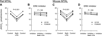
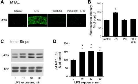
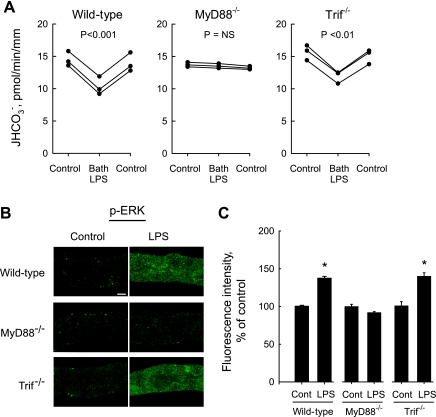
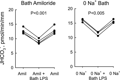
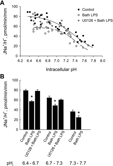
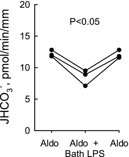
Sepsis is associated with defects in renal tubule function, but the underlying mechanisms are incompletely understood. Recently, we demonstrated that Gram-negative bacterial lipopolysaccharide (LPS) inhibits HCO(3)(-) absorption in the medullary thick ascending limb (MTAL) through activation of Toll-like receptor 4 (TLR4). Here, we examined the mechanisms responsible for inhibition of HCO(3)(-) absorption by basolateral LPS. Adding LPS to the bath decreased HCO(3)(-) absorption by 30% in rat and mouse MTALs perfused in vitro. The inhibition of HCO(3)(-) absorption was eliminated by the mitogen-activated protein kinase/extracellular signal-regulated kinase (MEK)/ERK inhibitors U0126 and PD98059. LPS induced a rapid (<15 min) and sustained (up to 60 min) increase in ERK phosphorylation in microdissected MTALs that was blocked by PD98059. The effects of basolateral LPS to activate ERK and inhibit HCO(3)(-) absorption were eliminated in MTALs from TLR4(-/-) and myeloid differentiation factor 88 (MyD88)(-/-) mice but were preserved in MTALs from TIR (Toll/interleukin-1 receptor) domain-containing adapter-inducing interferon-β (Trif)(-/-) mice. Basolateral LPS decreased apical Na(+)/H(+) exchanger 3 NHE3 activity through a decrease in maximal velocity (V(max)). The inhibition of NHE3 by LPS was eliminated by MEK/ERK inhibitors. LPS inhibited HCO(3)(-) absorption despite the presence of physiological stimuli that activate ERK in the MTAL. We conclude that basolateral LPS inhibits HCO(3)(-) absorption in the MTAL through activation of a TLR4/MyD88/MEK/ERK pathway coupled to inhibition of NHE3. These studies identify NHE3 as a target of TLR4 signaling in the MTAL and show that bacterial molecules can impair the absorptive functions of renal tubules through inhibition of this exchanger. The ERK pathway links TLR4 to downstream modulation of ion transport proteins and represents a potential target for treatment of sepsis-induced renal tubule dysfunction.


Figures

Fig. 1.
Fig. 1.
Inhibitors of extracellular signal-regulated kinase (ERK) activation eliminate inhibition of HCO3 absorption by bath lipopolysaccharide (LPS). Medullary thick ascending limbs (MTAL) from Sprague-Dawley rats and C57BL/6J mice were isolated and perfused in vitro. Tubules were studied in control solution (A and C) or bathed with 15 μM U0126 or 15 μM PD98059 (B and D), and then LPS (500 ng/ml) was added to and removed from the bath solution. Absolute rates of HCO3 absorption (JHCO3) were measured as described in methods. Data points are average values for single tubules. Lines connect paired measurements made in the same tubule. P values are for paired t-test. NS, not significant. Mean values are given in results.
Fig. 2.
Fig. 2.
LPS increases ERK phosphorylation in the MTAL. A: MTALs dissected from rats were incubated in vitro at 37°C in control solution, LPS (500 ng/ml), PD98059 (15 μM), or PD98059 + LPS for 15 min and then fixed and stained with antiphospho-ERK1/2-Thr202/Tyr204 (p-ERK) antibody. The tubules were analyzed by confocal immunofluorescence as described in methods. Images are Z-axis sections (0.4 μm) taken through a plane at the center of the tubule showing a cross-sectional view of cells in the lateral tubule walls (30, 31, 78). LPS increased p-ERK labeling, and this increase was eliminated by PD98059. Images are representative of at least eight tubules of each type. Scale bar = 5 μm. B: intensity of p-ERK staining for experiments in A was quantified as described in methods and is presented as a percentage of the control level. Bars are means ± SE. *P < 0.05 vs. control (ANOVA). C: time course of ERK activation. Inner stripe tissue from rats was incubated in vitro at 37°C in the absence and presence of LPS for the indicated times, and cell lysates were then immunoblotted with p-ERK antibody for analysis of ERK phosphorylation and anti-ERK antibody for total ERK level. Blots are representative of 3 independent experiments. D: p-ERK levels normalized for total ERK were determined for experiments in C by densitometry. p-ERK/ERK ratios are presented as a percentage of the control value (0 min) measured in the same experiment. Bars are means ± SE. *P < 0.05 vs. 0 min.
Fig. 3.
Fig. 3.
LPS-induced ERK activation is mediated through Toll-like recptor 4 (TLR4). A: MTALs dissected from wild-type and TLR4−/− mice were incubated in vitro for 15 min at 37°C in the absence (control) and presence of LPS. The tubules were then fixed and stained with p-ERK antibody and analyzed by confocal immunofluorescence as in Fig. 2A. LPS increased ERK phosphorylation in MTALs from wild-type mice but had no effect in MTALs from TLR4−/− mice. Images are representative of at least six tubules of each type. B: intensity of p-ERK staining was quantified for experiments in A as described in methods and is presented as a percentage of control level measured in the same experiment. *P < 0.05 vs. wild-type control.
Fig. 4.
Fig. 4.
Role of myeloid differentiation factor 88 (MyD88) and TIR (Toll/interleukin-1 receptor) domain-containing adapter-inducing interferon-β (Trif) in responses to LPS. A: MTALs from wild-type, MyD88−/−, and Trif−/− mice were perfused in vitro under control conditions, and then LPS (500 ng/ml) was added to and removed from the bath solution. JHCO3, data points, lines, and P values are as in Fig. 1. NS, not significant. B: MTALs from wild-type, MyD88−/−, and Trif−/− mice were incubated for 15 min at 37°C in vitro in the absence (control) and presence of LPS. The tubules were stained with p-ERK antibody and analyzed by confocal immunofluorescence as in Fig. 2A. The effect of LPS to increase ERK phosphorylation was eliminated in MTALs from MyD88−/− mice. Images are representative of at least six tubules of each type. C: intensity of p-ERK staining was quantified for experiments in B as described in methods and is presented as a percentage of control level measured in the same experiment. *P < 0.05 vs. control. Control fluorescence intensity did not differ in the three groups.
Fig. 5.
Fig. 5.
Inhibition of HCO3 absorption by bath LPS does not involve basolateral Na+/H+ exchange. MTALs from rats were studied with 10 μM amiloride in the bath (A) or in a Na+-free bath (B), conditions that inhibit basolateral Na+/H+ exchange (28, 34, 77, 78). LPS (500 ng/ml) was then added to and removed from the bath solution. In B, Na+ in the bath was replaced with NMDG+; the lumen was perfused with control solution containing 146 mM Na+ (28, 77). JHCO3, data points, lines, and P values are as in Fig. 1. Mean values are given in results.
Fig. 6.
Fig. 6.
Bath LPS inhibits apical Na+/H+ exchange through an ERK-dependent pathway. A: MTALs from rats were studied under control conditions and with 500 ng/ml LPS or 15 μM U0126 + LPS in the bath for 15–20 min. Apical Na+/H+ exchange rates (JNa+/H+) were determined from initial rates of pHi increase measured after addition of Na+ to the tubule lumen (see methods). Data points are from 16 control tubules, 19 tubules with LPS, and 10 tubules with U0126 + LPS. Solid (control) and dashed (bath LPS) lines are from least-squares fits to the Hill equation (81, 82); kinetic parameters (Vmax and apparent affinity for intracellular H+) are given in results. B: data from A were grouped over the indicated pHi intervals to obtain mean exchange rates (± SE). *P < 0.05 vs. other conditions in each interval. U0126 alone has no effect on apical Na+/H+ exchange activity (79).
Fig. 7.
Fig. 7.
Inhibition of HCO3 absorption by bath LPS is additive to inhibition by aldosterone. MTALs from rats were bathed with aldosterone (1 nM) and then LPS (500 ng/ml) was added to and removed from the bath solution. JHCO3, data points, lines, and P value are as in Fig. 1.
Fig. 8.
Fig. 8.
Model for inhibition of HCO3 absorption by basolateral LPS in the MTAL. Basolateral LPS decreases HCO3 absorption by inhibiting Na+/H+ exchanger 3 NHE3 through activation of the MEK/ERK signaling pathway. The LPS-induced ERK activation is mediated through TLR4 and the adaptor molecule MyD88. Arrows do not necessarily imply direct relationships; regulatory steps may involve additional signaling components.

Comment in

Similar articles

Cited by

References

    1. Alexander RT, Grinstein S. Tethering, recycling and activation of the epithelial sodium-proton exchanger, NHE-3. J Exp Biol 212: 1630–1637, 2009 - PubMed
    1. Alpern RJ. Renal acidification mechanisms. In: The Kidney, edited by Brenner BM, Rector FC., Jr Philadelphia, PA: Saunders, 2000, Vol. I, p. 455–519
    1. Amemiya M, Loffing J, Lotscher M, Kaissling B, Alpern RJ, Moe OW. Expression of NHE-3 in the apical membrane of rat renal proximal tubule and thick ascending limb. Kidney Int 48: 1206–1215, 1995 - PubMed
    1. Banerjee A, Gerondakis S. Coordinating TLR-activated signaling pathways in cells of the immune system. Immunol Cell Biol 85: 420–424, 2007 - PubMed
    1. Banerjee A, Gugasyan R, McMahon M, Gerondakis S. Diverse Toll-like receptors utilize Tpl2 to activate extracellular signal-regulated kinase (ERK) in hemopoietic cells. Proc Natl Acad Sci USA 103: 3274–3279, 2006 - PMC - PubMed

Publication types

MeSH terms