. 2011 Nov 3;118(18):5000-10.
doi: 10.1182/blood-2011-06-360743. Epub 2011 Aug 31.

Critical role for Syk in responses to vascular injury

Affiliations

Critical role for Syk in responses to vascular injury

Patrick Andre et al. Blood. .

Abstract

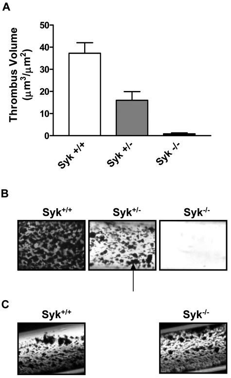
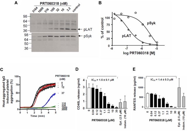
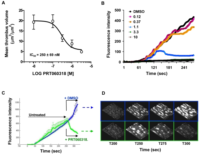
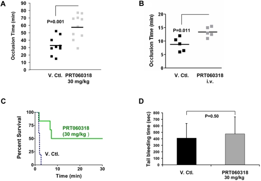
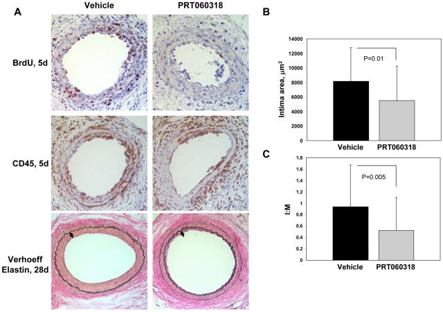
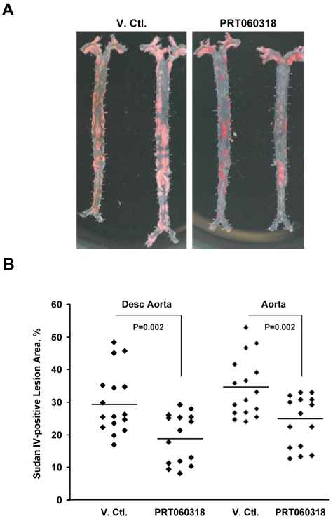
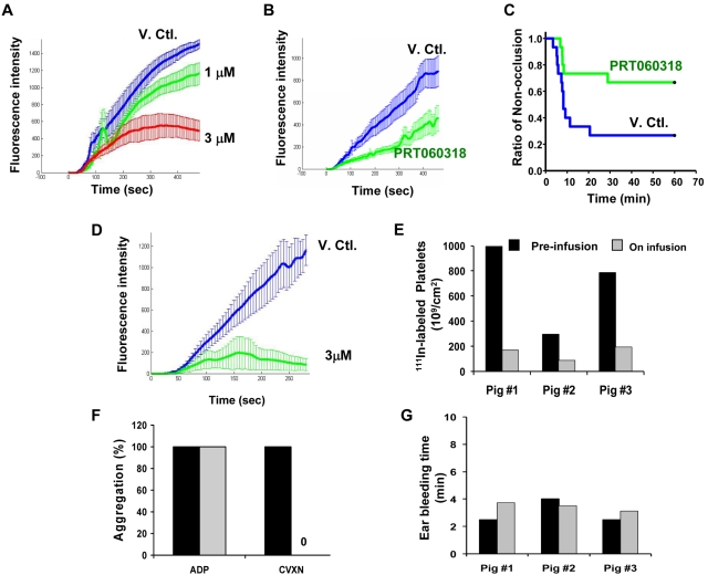
Although current antiplatelet therapies provide potent antithrombotic effects, their efficacy is limited by a heightened risk of bleeding and failure to affect vascular remodeling after injury. New lines of research suggest that thrombosis and hemorrhage may be uncoupled at the interface of pathways controlling thrombosis and inflammation. Here, as one remarkable example, studies using a novel and highly selective pharmacologic inhibitor of the spleen tyrosine kinase Syk [PRT060318; 2-((1R,2S)-2-aminocyclohexylamino)-4-(m-tolylamino)pyrimidine-5-carboxamide] coupled with genetic experiments, demonstrate that Syk inhibition ameliorates both the acute and chronic responses to vascular injury without affecting hemostasis. Specifically, lack of Syk (murine radiation chimeras) attenuated shear-induced thrombus formation ex vivo, and PRT060318 strongly inhibited arterial thrombosis in vivo in multiple animal species while having minimal impact on bleeding. Furthermore, leukocyte-platelet-dependent responses to vascular injury, including inflammatory cell recruitment and neointima formation, were markedly inhibited by PRT060318. Thus, Syk controls acute and long-term responses to arterial vascular injury. The therapeutic potential of Syk may be exemplary of a new class of antiatherothrombotic agents that target the interface between thrombosis and inflammation.


Figures

Figure 1
Figure 1
Lack of Syk attenuates arterial thrombus formation ex vivo. (A) Histogram showing the critical role for Syk in mediating thrombosis on type I collagen. (B) Thrombotic deposits formed on collagen-coated capillaries exposed for 2.5 minutes to nonanticoagulated blood from Syk+/+ (n = 9), Syk+/− (n = 10), and radiation chimeras (Syk−/− radiation chimera; n = 7) mice at 871 seconds−1. The black arrow points to platelet-rich thrombi. Quantification was performed on semithin cross section of Epon-embedded thrombotic deposits via computer-assisted morphometry. (C) Photomicrograph of thrombotic deposits formed on tissue factor-coated capillaries at 871 seconds−1 in Syk+/+ and Syk−/− radiation chimera mice.
Figure 2
Figure 2
Kinase specificity of PRT060318 and activity on human platelet function. (A) Integrilin-treated human washed platelets were incubated with 2 μL of various concentrations of PRT060318 (all in 30% DMSO) before activation with CVXN (150 ng/mL). Platelet lysates were subjected to SDS-PAGE gels, and Western blots were probed with anti-pLAT pY191 or pSyk pY525/526 antibodies. (B) Quantification of protein bands was performed using densitometry (Epson Expression 1680) using Quantity One Version 4.5.0 software (Bio-Rad Laboratories), and variations normalized to control bands (absence of PRT060318) were plotted against PRT060318 concentrations. (C) Representative dose response effect of PRT060318 on heat-aggregated IgG-induced platelet aggregation (n = 3; IC50 = 85nM in washed platelets). In PRP, PRT060318 inhibits collagen-induced platelet aggregation CD40L (D) and RANTES (E) release. Ept indicates eptifibatide; and Indom, indomethacin. For further data on PRT060318 specificity, please see Reilly et al.
Figure 3
Figure 3
Syk inhibition affects both thrombus initiation and thrombus stability in human blood. (A) Effect of PRT060318 on thrombus volume after perfusion of human blood over collagen. Each point represents the mean ± SEM of 7 individuals. (B) Continuous, real-time thrombosis profiles of 1 representative experiment in which anticoagulated human blood was perfused through collagen-coated capillaries in presence of PRT060318 (0.12-10μM). PRT060318 affects thrombus stability (< 1μM) and thrombus growth (> 3μM). (C) Mean thrombosis profiles of experiments performed with blood from 4 individuals showing dethrombotic activity of PRT060318. After 250 seconds, blood treated with either DMSO (vehicle control) or 5μM PRT060318 was immediately perfused over the preformed, untreated thrombi. (D) Representative 3-dimensional photomicrographs corresponding to panel C. The base of the thrombi remained unaffected by the treatment indicating destabilization of the platelet–platelet interactions.
Figure 4
Figure 4
PRT060318 inhibits arterial and venous thrombosis in the mouse. (A) PRT060318 (30 mg/kg oral) delays time to occlusion in carotid artery photochemical injury model (PRT060318, 58 ± 16 vs vehicle, 33 ± 12 minutes; P = .001). V.Ctl. indicates vehicle control. (B) PRT060318 (intravenous infusion) delays time to occlusion in FeCl3-injured mesenteric arteries (PRT060318, 13.4 ± 0.8 vs vehicle, 8.8 ± 1.1 minute; P = .011). (C) Inhibition of Syk by PRT060318 (30 mg/kg oral, n = 6) but not vehicle control (n = 5) prevents death after injection of a collagen + epinephrine mixture in WT mice. All vehicle control-treated animals died within 5 minutes of the intravenous. Injection, whereas 50% of the PRT060318-treated animals survived. (D) Tail bleeding times, performed by transecting the mouse tail 3 mm from the tip, are comparable in PRT060318 (30 mg/kg oral) and vehicle-treated mice (V. Ctl., n = 15; PRT060318, n = 14).
Figure 5
Figure 5
PRT060318 inhibits neointima formation after femoral artery wire injury. (A) Photomicrographs of injured femoral arteries from vehicle control and PRT060318-treated mice after wire injury. Verhoeff elastin staining 28 days after injury. Arrows indicate the internal elastic lamina. Images were captured using a microscope (model DM2000; Leica) and captured with an AxioCam MRc5 camera (Carl Zeiss) interfaced to a computer running Zeiss Axiovision Rel 4.5 software (original magnification, ×20). Immunostaining for CD45 and BrdU 5 days after injury. Quantitative morphometry, including intimal area (B) and intimal area:medial area (I:M) ratio (C), of mouse femoral arteries 28 days after injury.
Figure 6
Figure 6
Atherosclerotic lesion formation is attenuated by PRT060318. (A) Digital photograph (D70S camera [Nikon] with Tamron SP AF90mm F/2.8 Di Macro 1:1 lens) of Sudan IV staining of longitudinally opened and pinned thoraco-abdominal aortas harvested from ApoE−/− mice after 20 weeks of high-fat feeding. PRT060318 (30 mg/kg in distilled water; n = 15) or vehicle control (n = 16) were administered via oral gavage twice daily for 3 weeks followed by 1 week off, for a total of 16 weeks. (B) Quantification of percentage of lesion area in the descending thoracic and abdominal aorta as assessed by Sudan IV staining and computer-assisted imaging analysis.
Figure 7
Figure 7
PRT060318 activity in rabbits and pigs. (A) In vitro (spiking experiments) mean thrombotic profiles in presence of vehicle control (V.Ctl.) or PRT060318 (1 and 3μM) in rabbits. (B) Ex vivo thrombotic profiles associated with V. Ctl. or PRT060318 infusion regimen (intravenous infusion regimen was initiated before vascular injury and established as follows: from 0 to 15 minutes, 20.67 mg/kg/h at 12.4 mL/kg/h and then from 15 minutes until the end, 7.33 mg/kg/h at 4.4 mL/kg/h). (C) Occlusion rate in vivo in the rabbit thrombosis model. P = .021 by Gehan–Breslow survival analysis with Bonferroni comparison. (D) In vitro mean thrombotic profiles of whole blood from 4 pigs treated with DMSO (blue curve) or 3μM PRT060318. Intravenous infusion of PRT060318 (8.90 mg/kg/h at 1 mL/kg/h) in pigs inhibited 111In-labeled platelet deposition in vivo in the pig thrombosis model (E), abolished ex vivo platelet aggregation induced by CVXN (250 ng/mL) but not ADP (20μM; F), and did not affect the ear bleeding time (G).

References

    1. Libby P, Simon DI. Inflammation and thrombosis: the clot thickens. Circulation. 2001;103(13):1718–1720. - PubMed
    1. McEver RP. Adhesive interactions of leukocytes, platelets, and the vessel wall during hemostasis and inflammation. Thromb Haemost. 2001;86(3):746–756. - PubMed
    1. Massberg S, Brand K, Gruner S, et al. A critical role of platelet adhesion in the initiation of atherosclerotic lesion formation. J Exp Med. 2002;196(7):887–896. - PMC - PubMed
    1. Simon DI, Chen Z, Seifert P, Edelman ER, Ballantyne CM, Rogers C. Decreased neointimal formation in Mac-1(−/−) mice reveals a role for inflammation in vascular repair after angioplasty. J Clin Invest. 2000;105(3):293–300. - PMC - PubMed
    1. Palabrica T, Lobb R, Furie BC, et al. Leukocyte accumulation promoting fibrin deposition is mediated in vivo by P-selectin on adherent platelets. Nature. 1992;359(6398):848–851. - PubMed

Publication types

MeSH terms

Substances