. 2011 Oct 1;204(7):1086-94.
doi: 10.1093/infdis/jir467.

Lowering the threshold of lung innate immune cell activation alters susceptibility to secondary bacterial superinfection

Affiliations

Lowering the threshold of lung innate immune cell activation alters susceptibility to secondary bacterial superinfection

John Goulding et al. J Infect Dis. .

Abstract

Background: Previous studies have shown that the interaction of CD200R, a myeloid inhibitory receptor, with its ligand, CD200, is critical in the control of innate immune activation in the lung.

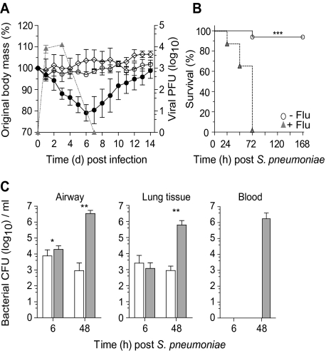
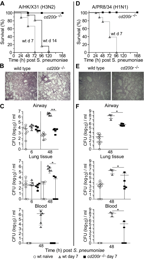
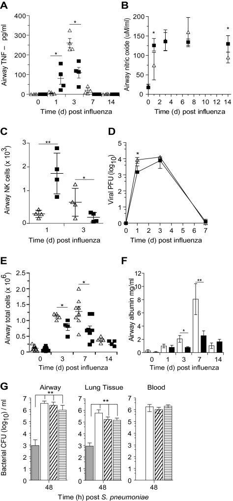
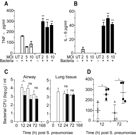
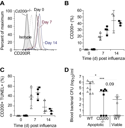
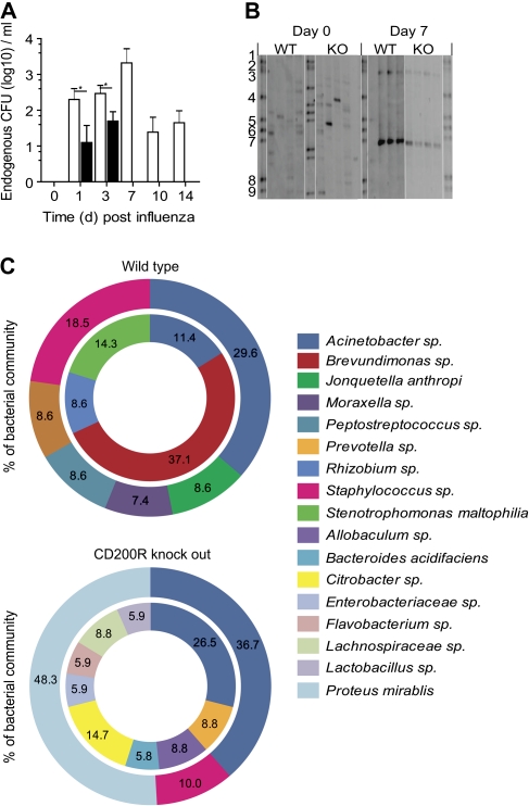
Methods and results: Using a mouse model of bacterial superinfection following influenza, we show that an absence of CD200R (a negative regulator highly expressed by macrophages and dendritic cells), restricts commensal and exogenous bacterial invasiveness and completely prevents the mortality observed in wild-type mice. This benefit is due to a heightened innate immune response to influenza virus in cd200r knockout mice that limits immune pathogenesis and viral load. In wild-type mice, apoptotic cells expressing CD200 that we believe contribute to the suppressed innate immune response to bacteria dominate during the resolution phase of influenza-induced inflammation. We also show for the first time the presence of a variety of previously unidentified bacterial species in the lower airways that are significantly adjusted by influenza virus infection and may contribute to the pathophysiology of disease.

Conclusions: The interaction of CD200 with CD200R therefore contributes to the hyporesponsive innate immune state following influenza virus infection that predisposes to secondary bacterial infection, a phenomenon that has the potential for immune modulation.


Figures

Figure 1.
Figure 1.
Influenza infection increases susceptibility to Streptococcus pneumoniae superinfection. A, Body mass (left axis) and viral titer (triangles, right axis) of wild-type C57BL/6 mice infected with 1.25 × 10⁵ plaque-forming units (PFU) of A/HK/X31 influenza (closed circle), 104 colony-forming units (CFU) Streptococcus pneumoniae (open circle), or vehicle control (diamonds). B, Survival of naive (open circle) or day 7 influenza-infected (triangles) wild-type mice infected with 104 CFU S. pneumoniae. C, Viable bacterial CFU recovered from the airway, lung tissue, and peripheral blood of naive (open bar) or day 7 postinfluenza (gray bar) mice at 6 hours and 48 hours following 104 CFU S. pneumoniae. Two-way ANOVA was used to interrogate weight loss, survival differences were determined using the log-rank Mantel–Cox test, and all other data were tested using a 2-tailed Mann–Whitney t test with 95% confidence intervals. Data are representative of 4 independent experiments (n = 5 mice per group). * P < .05, ** P < .01, *** P < .001 versus corresponding group.
Figure 2.
Figure 2.
Loss of CD200R prevents influenza-induced secondary bacterial superinfection. Survival of wild-type (wt) and cd200r−/− mice after infection with 104 colony-forming units (CFU) Streptococcus pneumoniae 7 days (gray triangles) or 14 days (open triangles) following A/HK/X31 (H3N2) (A) or A/PR8/34 (H1N1) (D) influenza virus infection. Representative hematoxylin and eosin–stained lung sections from day 7 A/X31 (H3N2) (B) or A/PR8/34 (H1N1) (E) influenza-infected wild-type and cd200r−/− mice 48 hours after S. pneumoniae infection. Total bacterial CFU recovered from the airway, lung tissue, and peripheral blood at 6 hours and 48 hours following 104 cfu S. pneumoniae in naive wild-type mice (circles) or those at day 7 of a A/HK/X31 (C) or A/PR8/34 (H1N1) (F) influenza infection. Survival differences were determined using the log-rank Mantel–Cox test, and all other data were tested using a 2-tailed Mann–Whitney t test with 95% confidence intervals. The data are representative of 3 independent experiments (n = 5–10 mice per group). * P < .05, ** P < .01, *** P < .001 versus corresponding group.
Figure 3.
Figure 3.
Antiviral immunity is enhanced in cd200r−/− mice. Airway levels of tumor necrosis factor (TNF)–α (A) and nitric oxide (B) at homeostasis (naive) and 1, 3, 7, and 14 days after infection with A/HK/X31 (H3N2) influenza virus in wild-type (triangles) and cd200r−/− (squares) mice. Natural killer (NK) cell number (C) and lung tissue viral titer (D) in wild-type (triangles) and cd200r−/− mice (squares) at 1, 3, and 7 days following A/HK/X31 (H3N2) influenza infection. Total airway cellularity (E) and albumin (F) following infection with A/HK/X31 (H3N2) influenza virus in wild-type (triangles, open bars) and cd200r−/− mice (squares, closed bars). G, Viable bacterial colony-forming units (CFU) recovered from the airway, lung tissue, and peripheral blood of naive wild-type mice (closed bar) or those at day 7 after infection with 1.25 × 10⁵ (open bar), 3.13 × 10⁴ (diagonal lines), or 2.08 × 10⁴ (horizontal lines) plaque-forming units (PFU) of influenza virus at 48 hours following 104 CFU Streptococcus pneumoniae. Cytokine data were analyzed using an unpaired 2-tailed Student t-test, assuming unequal variance. All other data were tested using a 2-tailed Mann–Whitney t test with 95% confidence intervals. Bonferroni correction was used for multiple comparisons. Data are representative of 4 independent experiments (n = 6–8 mice per group). * P < .05, ** P < .01, *** P < .001 versus corresponding group.
Figure 4.
Figure 4.
Enhanced bacterial responses in cd200r−/− mice. Tumor necrosis factor (TNF)–α (A) and interleukin (IL)–6 (B) liberated from wild-type (open bars) and cd200r−/− (closed bars) alveolar macrophages 24 hours following incubation with varying multiplicities of infection (MOI) of Streptococcus pneumoniae. C, Total colony-forming units (CFU) recovered from the airway and lung tissue of wild-type (open bar) and cd200r−/− mice (closed bar) at 12, 72, and 168 hours after 104 CFU S. pneumoniae infection. D, Airway levels of TNF-α in wild-type (triangles) and cd200r−/− mice (squares) 12 hours and 72 hours after S. pneumoniae infection. Cytokine data were analyzed using an unpaired 2-tailed Student t-test, assuming unequal variance. All other data were tested using a 2-tailed Mann–Whitney t test with 95% confidence intervals. Bonferroni correction was used for multiple comparisons. The data are representative of 4 independent experiments (n = 4 mice per group). * P < .05, ** P < .01, *** P < .001 versus corresponding group.
Figure 5.
Figure 5.
CD200 is expressed on apoptotic leukocytes in the airway during the resolution of influenza infection. A, Relative CD200R levels at day 0 (black), day 7 (red), and day 14 (blue) in wild-type (WT) mice compared with isotype matched control (dotted line) or staining of cd200r−/− airway macrophages (shaded). Percentage of CD200+ (B) and TUNEL+ CD200+ (C) airway monocytes/macrophages 0, 7, and 14 days after A/HK/X31 (H3N2) influenza infection. D, Intratracheal transfer of apoptotic WT (triangles), apoptotic cd200−/− (squares), or viable WT (circles) splenocytes into naive WT mice 16 hours prior to infection with 105 CFU Streptococcus pneumoniae. Data represents resultant blood colony-forming units (CFU) per mouse with mean ± SD after 48 hours. All data were tested using a 2-tailed Mann–Whitney t test with 95% confidence intervals. Bonferroni correction was used for multiple comparisons. The data are representative of 3 independent experiments (n = 6–8 per group). * P < .05, ** P < .01, *** P < .001 versus corresponding group.
Figure 6.
Figure 6.
Influenza infection causes a dysbiosis of the endogenous microbiota. A, Endogenous bacterial colony-forming units (CFU) recovered from the airway of wild-type (WT; open bar) and cd200r−/− (closed bar) mice following infection with A/HK/X31 (H3N2) influenza virus. Data were tested using a 2-tailed Mann–Whitney t test with 95% confidence intervals. Data are representative of 3 independent experiments (n = 5 mice per group). * P < .05, ** P < .01, *** P < .001 versus corresponding group. B, Denaturing gradient gel electrophoresis of bacterial 16s ribosomal RNA gene amplicon pools from the airways of naive and day 7 influenza-infected WT and cd200r−/− (KO) mice (n = 4 mice per group). Bands 1–9 represent a standardized bacteria species marker. C, Phylogenetic analysis of endogenous airway bacteria, expressed as a percentage of the total microbiota, from naive (inner circle) and day 7 influenza-infected (outer circle) WT and cd200r−/− mice. Only bacteria representing >5% or designating a known species name are shown. The omitted bacteria can be found in Supplemenatry Figure 1A. Data represent 2 independent samples randomly picked from each experimental group for cloning analysis and species identification.

References

    1. Brundage JF, Shanks GD. Deaths from bacterial pneumonia during 1918–19 influenza pandemic. Emerg Infect Dis. 2008;14:1193–9. - PMC - PubMed
    1. Centers for Disease Control and Prevention. Bacterial coinfections in lung tissue specimens from fatal cases of 2009 pandemic influenza A (H1N1)—United States, May–August 2009. MMWR Morb Mortal Wkly Rep. 2009;58:1071–4. - PubMed
    1. Cundell DR, Gerard NP, Gerard C, Idanpaan-Heikkila I, Tuomanen EI. Streptococcus pneumoniae anchor to activated human cells by the receptor for platelet-activating factor. Nature (London) 1995;377:435–8. - PubMed
    1. McCullers JA, Bartmess KC. Role of neuraminidase in lethal synergism between influenza virus and Streptococcus pneumoniae. J Infect Dis. 2003;187:1000–9. - PubMed
    1. Mandelboim O, Lieberman N, Lev M, et al. Recognition of haemagglutinins on virus-infected cells by NKp46 activates lysis by human NK cells. Nature (London) 2001;409:1055–60. - PubMed

Publication types

MeSH terms