. 2011 Oct;121(10):4150-60.
doi: 10.1172/JCI58414. Epub 2011 Sep 1.

A mutation in the immunoproteasome subunit PSMB8 causes autoinflammation and lipodystrophy in humans

Affiliations

A mutation in the immunoproteasome subunit PSMB8 causes autoinflammation and lipodystrophy in humans

Akiko Kitamura et al. J Clin Invest. 2011 Oct.

Abstract

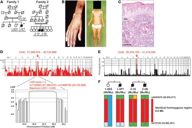
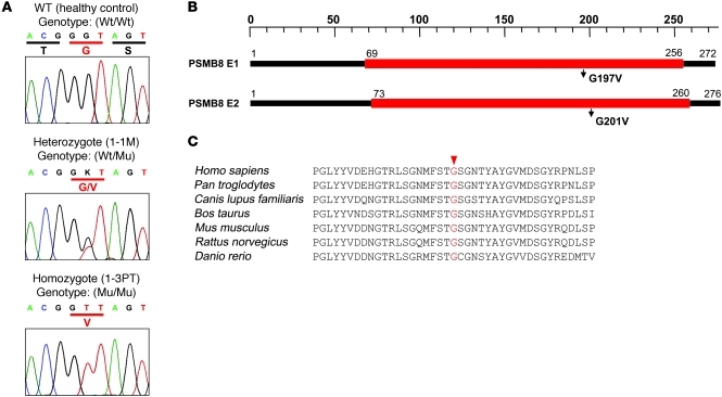
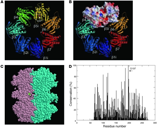
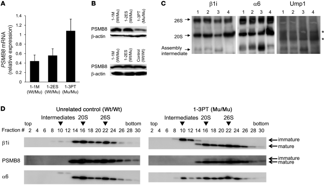
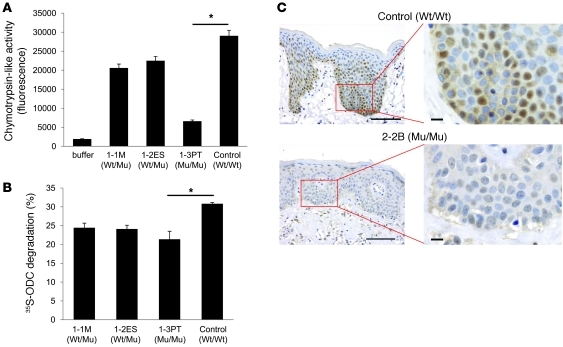
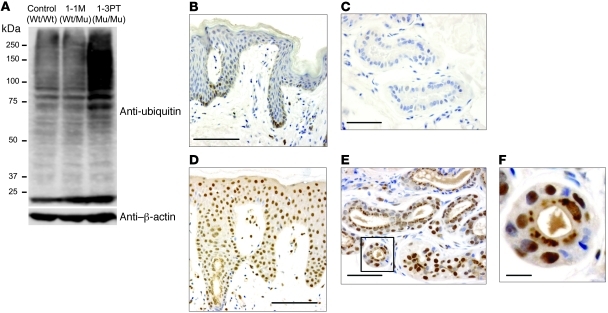
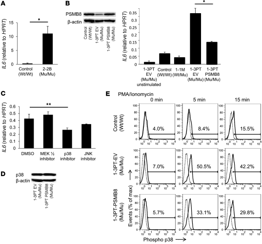
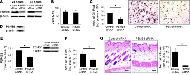
Proteasomes are multisubunit proteases that play a critical role in maintaining cellular function through the selective degradation of ubiquitinated proteins. When 3 additional β subunits, expression of which is induced by IFN-γ, are substituted for their constitutively expressed counterparts, the structure is converted to an immunoproteasome. However, the underlying roles of immunoproteasomes in human diseases are poorly understood. Using exome analysis, we found a homozygous missense mutation (G197V) in immunoproteasome subunit, β type 8 (PSMB8), which encodes one of the β subunits induced by IFN-γ in patients from 2 consanguineous families. Patients bearing this mutation suffered from autoinflammatory responses that included recurrent fever and nodular erythema together with lipodystrophy. This mutation increased assembly intermediates of immunoproteasomes, resulting in decreased proteasome function and ubiquitin-coupled protein accumulation in the patient's tissues. In the patient's skin and B cells, IL-6 was highly expressed, and there was reduced expression of PSMB8. Downregulation of PSMB8 inhibited the differentiation of murine and human adipocytes in vitro, and injection of siRNA against Psmb8 in mouse skin reduced adipocyte tissue volume. These findings identify PSMB8 as an essential component and regulator not only of inflammation, but also of adipocyte differentiation, and indicate that immunoproteasomes have pleiotropic functions in maintaining the homeostasis of a variety of cell types.


Figures

Figure 1
Figure 1. Pedigree structure and pathological features.
(A) Pedigree structures of 2 families. Black symbols denote patients with JASL; double horizontal lines denote consanguinity in a married couple; asterisks denote family members whose samples were obtained. Genotypes of PSMB8 are shown (Wt, wild-type allele; Mu, mutant allele). (B) Nodular erythema and deformity of fingers as well as partial lipodystrophy (face, upper body, distal parts of extremities) in 1-3PT. (C) Skin sections of 2-2B stained with hematoxylin and eosin. Original magnification, ×10 (top); ×20 (bottom). Scale bars: 100 μm. (D) Linkage analysis was performed using Merlin software; a linked region (rs1102563 [27,268,074] to rs10498750 [40,724,868]) with LOD score >3 on chromosome 6p (maximum LOD score, 3.435) is indicated by a red arrow. (E) Homozygosity mapping was carried out using HomozygosityMapper; homozygous regions are indicated in red. (F) Haplotype analysis around the homozygous region, carried out by SNP data, identified an identical homozygous region between rs9283878 and rs210120 (6.8 Mb; red) as a shared identical haplotype by descent in both families. PSMB8 genotypes are shown.
Figure 2
Figure 2. A missense mutation in PSMB8.
(A) Sequences obtained from a healthy control with wild-type PSMB8; from 1-1M, characteristic of a person with a heterozygous PSMB8 mutation, with a single nucleotide exchange (G→T) in exon 5 of PSMB8; and 1-3PT, characteristic of a homozygous PSMB8 mutation. PSMB8 genotypes are shown. (B) 2 isoforms of PSMB8 and the location of the mutation. (C) The amino acid sequence of human PSMB8 was compared with those of Pan troglodytes, Canis lupus familiaris, Bos taurus, Mus musculus, Rattus norvegicus, and Danio rerio. Conserved residues at the mutated positions are indicated in red.
Figure 3
Figure 3. Structure of PSMB8.
(A) The mouse subunits β1i, β2i, and β5i (i.e., PSMB8) were modeled by the corresponding constitutive subunits using Spanner software (see Methods). G197 is depicted in space-filling representation using CPK coloring. (B) β1i, β2i, and β5i were modeled by the corresponding constitutive subunits using Spanner software. Molecular surfaces of β4 and β5i were colored according to electrostatic potential: red, white, and blue represent negative, neutral, and positive electrostatic values, respectively. The location of the S1 substrate pocket (yellow circle) and the position of G197 (arrow) are indicated. (C) The cross-section of 2 β rings of PSMB8 (pink and cyan), shown using jV software ( http://www.pdbj.org/jv/index.html). G197 (green space-filling representation) was located outside the β ring–β ring interface. (D) Sequences of PSMB8 and 83 related proteins were obtained by running BLAST against the protein data bank. These sequences were aligned using MAFFT software, after removing several short fragments that did not cover the entire aligned region. The sequence conservation was computed and plotted, demonstrating that G197 was the most conserved position (100%) in the 84 related sequences.
Figure 4
Figure 4. Defective immunoproteasome complexes in JASL cells.
(A) PSMB8 mRNA expression in transformed B cells from 1-1M, 1-2ES, and 1-3PT were evaluated by real-time PCR. Data are representative of 4 experiments. (B) Expression of PSMB8 and β-actin in EBV-transformed B cells from 1-1M, 1-2ES, 1-3PT, and an unrelated control, evaluated by Western blotting. (C) Cell lysates from transformed B cells of 1-1M (lane 1), 1-2ES (lane 2), 1-3PT (lane 3), and unrelated healthy control (lane 4) were subjected to native PAGE and immunoblotted with anti-Ump1, anti-α6, and anti-β1i polyclonal antibodies. Arrows denote bands corresponding to the 20S or 26S proteasome, as well as intermediate complexes. Asterisks denote nonspecific bands. (D) Glycerol gradient centrifugation was performed by using extracts of transformed B cells from 1-3PT and an unrelated healthy control. Fractions were immunoblotted with anti-α6, anti-β1i, and anti-PSMB8 antibody. Arrowheads denote assembly intermediates as well as 20S and 26S immunoproteasomes. PSMB8 genotypes are shown. All data are representative of at least 4 experiments.
Figure 5
Figure 5. A mutation in PSMB8 reduces proteasomes activity.
(A) Chymotrypsin-like activities of 26S proteasomes in transformed B cells from 1-3PT, 1-1M, 1-2ES, and unrelated control. *P < 0.01. (B) Cell extracts from transformed B cells were assayed for adenosine triphosphate–dependent protein degradation activity using 35S-labeled ODC. *P < 0.01. (C) Skin sections from a healthy control and 2-2B were stained with anti-PSMB8 mAb and hematoxylin. Scale bars: 100 μm (left); 10 μm (right). PSMB8 genotypes are shown. All data are representative of at least 4 experiments.
Figure 6
Figure 6. Ubiquitinated proteins accumulate in JASL cells.
(A) Expression of ubiquitin in transformed B cells from healthy control, 1-1M, and 1-3PT, evaluated by Western blotting with anti-ubiquitin and β-actin mAbs. Data are representative of 5 experiments. (BF) Skin sections of a healthy control (B and C) and 2-2B (DF) stained with anti-ubiquitin mAb together with hematoxylin. Original magnification, ×20 (B and D); ×40 (C and E). The boxed region in E is shown enlarged in F. Histological data are representative of 4 independent staining experiments using a biopsy sample from each patient. Scale bars: 100 μm (B and D); 50 μm (C and E); 10 μm (F).
Figure 7
Figure 7. A missense mutation in PSMB8 hyperactivates B cells.
(A) IL6 expression in skin from a healthy donor and 2-2B, evaluated by real-time PCR. *P < 0.01. (B) Transformed B cells from 1-3PT were transduced with PSMB8 or a control vector. Expression of PSMB8 in healthy control, 1-3PT–EV, and 1-3PT–PSMB8 transformed B cells were evaluated by Western blotting. Expression of IL6 in PMA- and ionomycin-stimulated control, 1-1M, 1-3PT–EV, and 1-3PT–PSMB8 B cells were evaluated by real-time PCR. *P < 0.01. (C) Expression of IL6 in cells after PMA and ionomycin stimulation in the presence of MEK1/2, p38, or JNK inhibitors, evaluated by real-time PCR. **P < 0.05. (D) Expression of total p38 in 1-3PT–EV and 1-3PT–PSMB8 cells, evaluated by Western blot. (E) Phospho-p38 in healthy control, 1-3PT–EV, and 1-3PT–PSMB8 transformed B cells after PMA and ionomycin stimulation. Gray and black histograms denote cells stained with control rabbit IgG and anti–phospho-p38, respectively. Percentages within histograms denote expression of phospho-p38 in PMA- and ionomycin-stimulated cells. All data are representative of 7 experiments.
Figure 8
Figure 8. A missense mutation in PSMB8 blocks adipocyte differentiation.
(A and B) 3T3-L1 cells were transfected with siRNA against PSMB8 or control siRNA. (A) Expression of PSMB8 was evaluated 24 and 48 hours after transfection by Western blotting. (B) Cell viability was tested by trypan blue staining 48 hours after transfection. (C) Induction of adipocytes was evaluated by staining cells with Oil Red 6 days after induction, and Oil Red–positive cells were also quantified. *P < 0.01. (D) Expression of PSMB8 in human preadipocytes was examined by Western blotting. (E) Expression of PSMB8 or β-actin in human preadipocytes transfected with siRNA against PSMB8 or control siRNA (24 hours after transfection) was evaluated by Western blotting and quantified. *P < 0.01. (F) Human preadipocytes were transfected with siRNA against PSMB8 and induced to differentiate into adipocytes; 12 days after transfection, Oil Red–positive cells were evaluated by densitometry. *P < 0.01. (G) siRNA for PSMB8 or control siRNA was subcutaneously injected into the skin of BALB/c mice. The skin was obtained 11 days after the initial siRNA injection, and the section was stained with hematoxylin and eosin. Original magnification, ×10. Scale bars: 100 μm. Moreover, the number of hair follicles in 10,000 μm2 (10 regions) was counted. *P < 0.01. All data are representative of at least 4 experiments.

References

    1. Masters SL, Lobito AA, Chae J, Kastner DL. Recent advances in the molecular pathogenesis of hereditary recurrent fevers. Curr Opin Allergy Clin Immunol. 2006;6(6):428–433. doi: 10.1097/ACI.0b013e3280109b57. - DOI - PubMed
    1. Dinarello CA. Interleukin-1beta and the autoinflammatory diseases. N Engl J Med. 2009;360(23):2467–2470. - PubMed
    1. Dinarello CA. Immunological and inflammatory functions of the interleukin-1 family. Annu Rev Immunol. 2009;27:519–550. doi: 10.1146/annurev.immunol.021908.132612. - DOI - PubMed
    1. McDermott MF, et al. Germline mutations in the extracellular domains of the 55 kDa TNF receptor, TNFR1, define a family of dominantly inherited autoinflammatory syndromes. Cell. 1999;97(1):133–144. doi: 10.1016/S0092-8674(00)80721-7. - DOI - PubMed
    1. Finley D. Recognition and processing of ubiquitin-protein conjugates by the proteasome. Annu Rev Biochem. 2009;78:477–513. doi: 10.1146/annurev.biochem.78.081507.101607. - DOI - PMC - PubMed

Publication types

MeSH terms