. 2011 Oct;121(10):4030-42.
doi: 10.1172/JCI45114. Epub 2011 Sep 1.

Smad4 deficiency in T cells leads to the Th17-associated development of premalignant gastroduodenal lesions in mice

Affiliations

Smad4 deficiency in T cells leads to the Th17-associated development of premalignant gastroduodenal lesions in mice

Jennifer Nancy Hahn et al. J Clin Invest. 2011 Oct.

Abstract

While there is evidence that specific T cell populations can promote the growth of established tumors, instances where T cell activity causes neoplasms to arise de novo are infrequent. Here, we employed two conditional mutagenesis systems to delete the TGF-β signaling pathway component Smad4 in T cells and observed the spontaneous development of massive polyps within the gastroduodenal regions of mice. The epithelial lesions contained increased levels of transcripts encoding IL-11, IL-6, TGF-β, IL-1β, and TNF-α, and lamina propria cells isolated from lesions contained abundant IL-17A+CD4+ T cells. Furthermore, we found that Smad4 deficiency attenuated TGF-β-mediated in vitro polarization of FoxP3+CD4+ T cells, but not IL-17A+CD4+ T cells, suggesting that the epithelial lesions may have arisen as a consequence of unchecked Th17 cell activity. Proinflammatory cytokine production likely accounted for the raised levels of IL-11, a cytokine known to promote gastric epithelial cell survival and hyperplasia. Consistent with IL-11 having a pathogenic role in this model, we found evidence of Stat3 activation in the gastric polyps. Thus, our data indicate that a chronic increase in gut Th17 cell activity can be associated with the development of premalignant lesions of the gastroduodenal region.


Figures
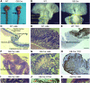
Figure 1
Figure 1. Hyperplastic gastric polyps in GB-Cre;Smad4fl/fl mice.
(A) The upper digestive tract of 12- to 18-month-old Smad4fl/fl (WT) and GB-Cre;Smad4fl/fl (GB-Cre) mice, with the latter showing swelling at the gastroduodenal junction (arrows). Scale bar: 0.5 cm. (B) A representative GB-Cre antro-pyloric polyp (dash-lined box) and WT antro-pyloric region. Scale bars: 0.25 cm. (CE) Antro-pyloric region of a WT mouse showing H&E staining and anti-TFF1 antibody reactivity (scale bars: 1,000 μm, 125 μm, and 500 μm, respectively). The pyloric junction (indicated by a dash-lined box in C), as well as a higher-magnification view of the antro-pyloric mucosa (D). Anti-TFF1 staining was limited to the gastric mucosa (E; arrow). (FK) GB-Cre gastric lesion sections stained with H&E, anti-TFF1 antibody, or anti-PCNA antibody (scale bars: 1,000 μm [F], 250 μm [G], 500 μm [H], 40 μm [IK]). A GB-Cre gastric polyp (F), with glands demonstrating elongation, branching, and dilation (G), as well as “side buds” (I; asterisks) containing abundant anti-PCNA antibody–reactive cells (J; black arrow indicates non-proliferating elongated gland; red arrows indicate proliferative side buds). Low-grade epithelial dysplasia was evident (K; arrows), as well as diffuse anti-TFF1 antibody staining of the polyp mucosa (H). Images are representative of 4–6 mice per group.
Figure 2
Figure 2. Lck-Cre;Smad4fl/fl mice develop proximal duodenal adenomas.
(AC) Proximal duodenal region of a 12-month-old WT (Smad4fl/fl) mouse (A, scale bar: 0.25 cm) showing normal arrangement of villi (B), and anti-PCNA antibody staining limited to the basal region of villous crypts (C) (scale bars: 250 μm). (DH) Representative duodenal adenomas in a 12-month-old Lck-Cre;Smad4fl/fl (Lck-Cre) mouse (D; arrows), located immediately distal to the pyloric junction (scale bar: 0.25 cm). The lesions showed elongated distended villi with a crowded glandular architecture (E; bracket) (scale bar: 250 μm), evidence of hematopoietic cell infiltrates (G) (scale bar: 250 μm), and areas of epithelial dysplasia (H, arrows) exhibiting a crowded and abnormal arrangement of nuclei (scale bar: 60 μm). (F) A representative section from an adenoma showing diffuse anti-PCNA antibody staining (scale bar: 250 μm). Images are a representative of 4 control and 4 experimental mice.
Figure 3
Figure 3. Characterization of GB-Cre–mediated Cre reporter gene activation.
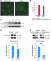
(A) Anti-EYFP antibody staining of a representative (of 3 mice) region of duodenum and the antro-pyloric polyp of an 18-month-old GB-Cre;Smad4fl/fl;R26R-EYFP mouse. EYFP immunostaining was present in lamina propria and intraepithelial inflammatory cells; however, the epithelium was negative for reporter gene expression (scale bars: 250 μm). (B) Flow cytometric detection of EYFP expression in 7-week and 56- to 60-week-old GB-Cre;R26R-EYFP splenocytes stained with anti-CD4 and anti-CD8 antibodies (data are shown as the mean ± SEM; n = 4 mice/group). Representative flow cytometry plots are shown in Supplemental Figure 2A. (C) Excision of Smad4 analyzed by PCR in 12- to 18-month-old Smad4fl/fl (WT) and GB-Cre;Smad4fl/fl (GB-Cre) CD8+ or CD4+ splenocytes (representative of 3 mice). (D) Immunoblotting of splenic T cells isolated from 12- to 18-month-old Smad4fl/fl and experimental (GB-Cre;Smad4fl/fl) mice for Smad4 expression levels, along with densitometry results (data shown are the mean ± SEM; n = 3 mice/group). (E) Immunoblotting for Smad4 expression in lysates from GB-Cre;Smad4fl/fl and Lck-Cre;Smad4fl/fl (Lck-Cre) splenic T cells following anti-CD3–mediated activation and expansion in IL-2 for 7–9 days, with densitometry (data are shown as the mean ± SEM; n = 3–4 mice/group). *P < 0.05, ***P < 0.001, 2-tailed unpaired t test.
Figure 4
Figure 4. Expression of two IL-6 family cytokines and evidence of Stat3 activation in GB-Cre;Smad4fl/fl antro-pyloric polyps.
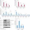
(A and C) Quantitative RT-PCR (qRT-PCR) real-time analysis of the cytokine transcripts from the antro-pyloric regions of 12- to 18-month-old Smad4fl/fl (WT), and polyp-containing GB-Cre;Smad4fl/fl (GB-Cre) mice. Data are depicted as the mean ± SEM (n = 4–5 mice/group). (B) qRT-PCR real-time analysis of cytokine transcripts from the proximal duodenum region of 12- to 18-month-old Smad4fl/fl and Lck-Cre;Smad4fl/fl (Lck-Cre) mice. Data are shown as the mean ± SEM (n = 4 mice/group). ActβA and ActβB, activin subunits βA and βB, respectively. (D) Immunoblotting of lysates prepared from the antro-pyloric region of 12- to 18-month-old control (WT) and polyp-containing GB-Cre;Smad4fl/fl mice, showing the expression levels of Stat3, phospho-Stat3, Stat1, and phospho-Stat1 proteins, with densitometry for phospho-Stat3 being normalized to Stat3 levels and phospho-Stat1 normalized to Stat1 levels. Data are shown as the mean ± SEM (n = 3–4 mice/group). *P < 0.05, **P < 0.01, ***P < 0.001, 2-tailed unpaired t test.
Figure 5
Figure 5. IL-17A expression in CD4+ T cells isolated from the lamina propria of GB-Cre;Smad4fl/fl polyps.
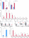
(A) Cell surface phenotyping of lamina propria cells isolated from the antro-pyloric regions of 12- to 18-month-old polyp-bearing GB-Cre;Smad4fl/fl mice and age-matched controls. Data are shown as the mean ± SEM (n = 4–5 mice/group). (B) qRT-PCR real-time analysis of transcripts present within polyp and control gut lamina propria cell RNA samples. Data are shown as the mean ± SEM (n = 3–4 mice/group). (CE) Intracellular staining for IL-17A within lamina propria cells from control and polyp-bearing mice. Samples were also stained for CD8+ (C) or for both CD4+ and CD11b+ (D) cells, and results for IL-17A+CD4+ cells are also shown (E). Data are shown as the mean ± SEM (n = 3 mice/group). (F) qRT-PCR real-time analysis of the indicated transcripts within RNA samples of lamina propria cells isolated from the proximal duodenum regions of 12- to 18-month-old experimental (Lck-Cre) and control (WT) mice. Data are shown as the mean ± SEM (n = 3–4 mice/group). *P < 0.05, **P < 0.01, ***P < 0.001, 2-tailed unpaired t test.
Figure 6
Figure 6. Smad4 deficiency impairs iTreg polarization.
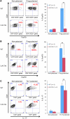
Splenic T cells from control (WT) and Smad4-deficient (Lck-Cre) mice were activated under non-polarizing (using plate-bound anti-CD3, anti-CD28) or iTreg-polarizing conditions (A). Naive T cells isolated from splenocytes from control (WT) and Smad4-deficient (Lck-Cre) mice were similarly activated under non-polarizing, iTreg-polarizing (B), or Th17-polarizing (C) conditions. FoxP3+ (A and B) or IL-17+ (C) expression was detected by intracellular cytokine staining and flow cytometry. Average results indicating the numbers of CD4+ T cells that were Foxp3+ (A and B) or IL-17A+ (C) are shown; data are represented as the mean ± SEM (n = 3–4 mice/group). **P < 0.01, 2-tailed unpaired t test.
Figure 7
Figure 7. IL-17A stimulation of gastric explants and stromal cell lines.
(A) qRT-PCR real-time analysis of the indicated transcripts of explanted tissue samples from the antro-pyloric regions of wild-type mice, cultured in either the presence or absence (unstim) of IL-17A. Data are the mean ± SEM (n = 3 mice/group). (B) qRT-PCR real-time analysis of Il11 and Il6 transcripts in M2-10B4 and D1 murine bone marrow stromal cell line RNA in response to IL-17A stimulation (n = 4–3). As a positive control, TGF-β stimulation (n = 5–6) was used. Data are the mean ± SEM. *P < 0.05, **P < 0.01, ***P < 0.001, 2-tailed unpaired t test.

Similar articles

Cited by

References

    1. Shull MM, et al. Targeted disruption of the mouse transforming growth factor-beta 1 gene results in multifocal inflammatory disease. Nature. 1992;359(6397):693–699. doi: 10.1038/359693a0. - DOI - PMC - PubMed
    1. Kulkarni AB, et al. Transforming growth factor-beta 1 null mice. An animal model for inflammatory disorders. Am J Pathol. 1995;146(1):264–275. - PMC - PubMed
    1. Kulkarni AB, et al. Transforming growth factor beta 1 null mutation in mice causes excessive inflammatory response and early death. Proc Natl Acad Sci U S A. 1993;90(2):770–774. doi: 10.1073/pnas.90.2.770. - DOI - PMC - PubMed
    1. Liu Y, Zhang P, Li J, Kulkarni AB, Perruche S, Chen W. A critical function for TGF-beta signaling in the development of natural CD4+CD25+Foxp3+ regulatory T cells. . Nat Immunol. 2008;9(6):632–640. doi: 10.1038/ni.1607. - DOI - PubMed
    1. Gorelik L, Flavell RA. Abrogation of TGFbeta signaling in T cells leads to spontaneous T cell differentiation and autoimmune disease. Immunity. 2000;12(2):171–181. doi: 10.1016/S1074-7613(00)80170-3. - DOI - PubMed

Publication types

MeSH terms