. 2011 Oct;121(10):4082-94.
doi: 10.1172/JCI57398. Epub 2011 Sep 1.

A sodium channel knockin mutant (NaV1.4-R669H) mouse model of hypokalemic periodic paralysis

Affiliations

A sodium channel knockin mutant (NaV1.4-R669H) mouse model of hypokalemic periodic paralysis

Fenfen Wu et al. J Clin Invest. 2011 Oct.

Abstract

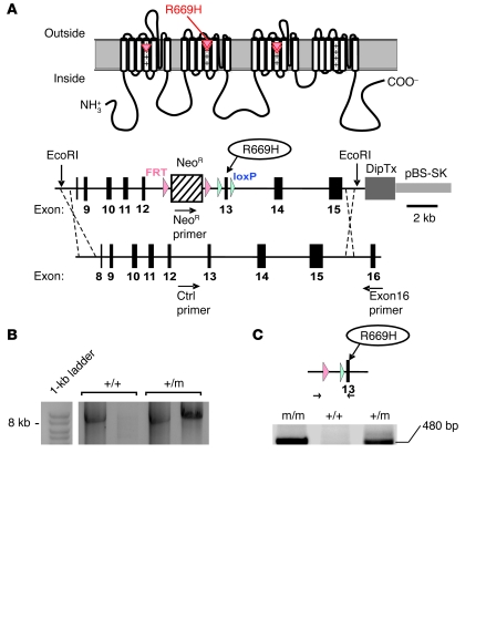
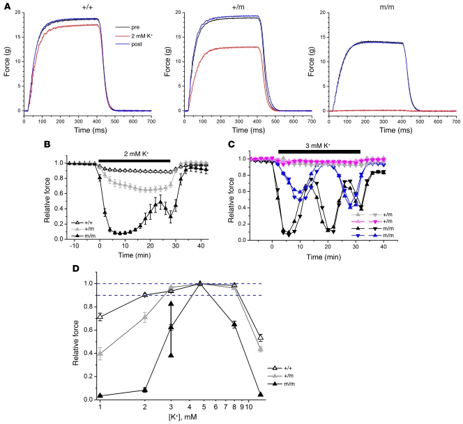
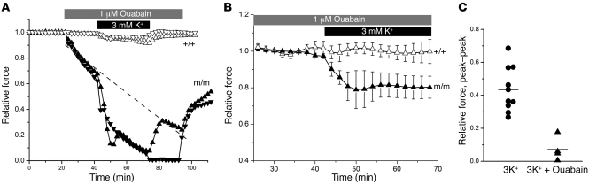
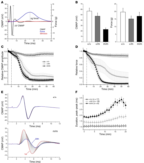
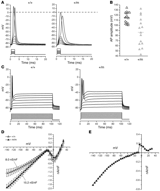
Hypokalemic periodic paralysis (HypoPP) is an ion channelopathy of skeletal muscle characterized by attacks of muscle weakness associated with low serum K+. HypoPP results from a transient failure of muscle fiber excitability. Mutations in the genes encoding a calcium channel (CaV1.1) and a sodium channel (NaV1.4) have been identified in HypoPP families. Mutations of NaV1.4 give rise to a heterogeneous group of muscle disorders, with gain-of-function defects causing myotonia or hyperkalemic periodic paralysis. To address the question of specificity for the allele encoding the NaV1.4-R669H variant as a cause of HypoPP and to produce a model system in which to characterize functional defects of the mutant channel and susceptibility to paralysis, we generated knockin mice carrying the ortholog of the gene encoding the NaV1.4-R669H variant (referred to herein as R669H mice). Homozygous R669H mice had a robust HypoPP phenotype, with transient loss of muscle excitability and weakness in low-K+ challenge, insensitivity to high-K+ challenge, dominant inheritance, and absence of myotonia. Recovery was sensitive to the Na+/K+-ATPase pump inhibitor ouabain. Affected fibers had an anomalous inward current at hyperpolarized potentials, consistent with the proposal that a leaky gating pore in R669H channels triggers attacks, whereas a reduction in the amplitude of action potentials implies additional loss-of-function changes for the mutant NaV1.4 channels.


Figures

Figure 1
Figure 1. Construction and genetic analysis of the NaV1.4-R669H knockin mouse.
(A) Schematic diagram of NaV1.4 showing the locations of the HypoPP mutations at arginines in S4 voltage sensor domains (red triangles). The targeting construct (22.4 kb) for homologous recombination in mouse SCN4A is shown below (see Methods for details). (B) PCR amplification of an 8.5-kb fragment from splenic DNA isolated from F1 mice showed integration upstream of exon 16 in SCN4A. The reverse primer was in exon 16, downstream from the targeting construct, as shown in A. The NeoR and exon 16 primer pair specifically amplified the R669H+/m allele containing the Neo cassette, but not the WT allele. (C) PCR analysis of genomic DNA, after crossing with a line expressing Flp recombinase, showed the removal of the Neo cassette with retention of 1 FRT site and 1 loxP site upstream of exon 13 (480 bp, compared with 400 bp for WT). The PCR products also demonstrated a homozygous mutant lacking the 400-bp amplimer (R669Hm/m). (D) RT-PCR from muscle total RNA using 40 cycles of amplification showed the specificity of forward primers to amplify only the WT allele R or the mutant allele H. (E) Expression levels of WT and mutant alleles were ascertained by RT-PCR for 24 cycles with allele-specific primers, with normalization to expression of the β-actin transcript. Expression of the mutant allele in R669Hm/m mice was comparable to that of the WT allele in WT mice, and each allele was present at approximately 70% of total control for R669H+/m mice.
Figure 2
Figure 2. In vitro contraction testing demonstrates a HypoPP phenotype.
(A) Force tracings of isometric tetanic contractions recorded in standard bath solution (4.75 mM K+; black), after 10 minutes in 2 mM K+ (red), and after recovering in standard solution for 10 minutes (blue). R669H+/m and R669Hm/m mice were more susceptible to hypokalemic-induced weakness. The baseline tetanic force was consistently lower in R669Hm/m mice. (B) Average responses for a 30-minute exposure to 2 mM K+ challenge (n = 10 [WT]; 8 [R669H+/m and R669Hm/m]). Tetanic force was recorded every 2 minutes and for each muscle and was normalized to the control response preceding the hypokalemic challenge. (C) Response to 3 mM K+ challenge, presented as paired recordings from individual soleus muscles from the left and right hindlimbs (tested in separate tissue baths). Paired muscles from the same animal are shown by symbol color. Large-amplitude oscillations in force were observed for all R669Hm/m muscles tested (n = 10). The pair of recordings from 2 different R669Hm/m mice illustrates the highly synchronous responses for muscles harvested from the same animal. (D) Dose-response relation for tetanic contraction after a 10-minute exposure to varying levels of K+. Average maximum and minimum forces observed during 30 minutes’ exposure are also indicated (vertical lines). For R669H+/m mice, increased susceptibility to weakness was observed at low K+, but not for high K+ as occurs in HyperPP. Dashed lines span the top 10% of relative force.
Figure 3
Figure 3. Recovery from weakness was ouabain sensitive.
In vitro contraction responses from R669Hm/m muscle exposed to 3 mM K+ were used to assess whether spontaneous recovery of force during hypokalemia was ouabain sensitive. (A) Pretreatment with 1 μM ouabain caused a decline in force for R669Hm/m, but not WT, muscle. Concurrent exposure to 3 mM K+ produced a further decline in force and suppressed large oscillation in force. Responses are from 2 individual soleus muscles harvested from R669Hm/m or WT mice. (B) Compensation for the ouabain-induced decline in force (dashed line in A) revealed the persistence of hypokalemia-induced weakness, but with suppression of spontaneous recovery. Responses are averages (n = 4 [R669Hm/m]; 2 [WT]). (C) Peak-to-peak amplitude for the spontaneous oscillation in R669Hm/m soleus force during 3 mM K+ challenge, shown for 10 separate muscles without ouabain and for 4 muscles pretreated with 1 μM ouabain.
Figure 4
Figure 4. In vivo reduction of muscle excitability and force from glucose plus insulin challenge.
(A) CMAP (black) and force (blue) at the Achilles tendon were recorded simultaneously in response to a single 0.1-ms shock (red) applied to the sciatic nerve. Sample tracings are from a single trial (nonaveraged) recorded from a R669H+/m mouse. (B) Baseline CMAP amplitude and force, recorded before glucose plus insulin infusion. Relative change in CMAP amplitude (C) and twitch force (D) in response to glucose and insulin infusion. Amplitudes were normalized to the average of 5 trials before the start of the infusion (vertical dashed lines). (E) Individual CMAP responses are superimposed for the first 20 responses, measured at 1-minute intervals, after glucose plus insulin infusion for WT and R669Hm/m animals. (F) The duration of the CMAP (peak to peak) was prolonged for R669H mutants and increased during glucose plus insulin infusion. (BD) Responses are averaged (n = 16 [WT]; 8 [R669H+/m]; 7 [R669Hm/m]).
Figure 5
Figure 5. Reduced muscle excitability in R669H fibers.
(A) Muscle voltage responses elicited by a series of 2-ms stimuli at progressively larger current amplitudes. A holding current was applied to set the initial membrane potential at –85 mV. Action potentials peaked with an overshoot greater than 0 mV (dashed line) for WT fibers, but not for all R669H+/m fibers. Maximal current stimulus was 1.5× threshold. (B) Action potential (AP) amplitudes elicited by 1.5× threshold stimuli were smaller for R669H+/m than WT fibers (n = 15 fibers from 5 muscle preparations per genotype). (C) Voltage transients in response to 100-ms stimulating current injections. A single action potential was elicited in both WT and R669H+/m fibers, even for stimulus currents of 1.5× threshold, demonstrating an absence of myotonia. (D) Voltage dependence for the steady-state current recorded at the end of a 300-ms voltage pulse revealed an increased inward (negative) current for R669Hm/m fibers. Currents are the lanthanum-sensitive component determined by subtraction of those recorded in a bath containing 3.5 mM La3+ from control responses. Inflection between –30 mV and +20 mV reflects a residual Ca2+ current blocked by La3+. (E) Subtraction of La-sensitive currents in WT fibers from R669Hm/m yielded the gating pore current that was activated at hyperpolarized potentials.
Figure 6
Figure 6. Histological analysis of quadriceps muscles.
Frozen blocks were sectioned at 10-μm thicknesses and stained with H&E (A, E, and I), Gomori trichrome (B, F, and J), nicotinamide adenine dehydrogenase-tetrazolium reductase (NADH-TR; C, G, and K), and myoadenylate deaminase (MADM; D, H, and L). Red-staining subsarcolemmal inclusions on Gomori trichrome (F and J) were the only changes observed for R669H mutant muscle. Scale bar: 50 μm.

Similar articles

Cited by

References

    1. Lehmann–Horn F, Rüdel R, Jurkat–Rott K. Nondystrophic myotonias and periodic paralyses. In: Engel AG, Franzini-Armstrong C, eds.Myology . New York, New York, USA: McGraw Hil; 2004:1257–1300.
    1. Cannon SC. Pathomechanisms in channelopathies of skeletal muscle and brain. Annu Rev Neurosci. 2006;29:387–415. doi: 10.1146/annurev.neuro.29.051605.112815. - DOI - PubMed
    1. Venance SL, et al. The primary periodic paralyses: diagnosis, pathogenesis and treatment. Brain. 2006;129(pt 1):8–17. doi: 10.1093/brain/awh639. - DOI - PubMed
    1. Rüdel R, Lehmann-Horn F, Ricker K, Kuther G. Hypokalemic periodic paralysis: in vitro investigation of muscle fiber membrane parameters. Muscle Nerve. 1984;7(2):110–120. doi: 10.1002/mus.880070205. - DOI - PubMed
    1. Elbaz A, et al. Hypokalemic periodic paralysis and the dihydropyridine receptor (CACNL1A3): genotype/phenotype correlations for two predominant mutations and evidence for the absence of a founder effect in 16 caucasian families. Am J Hum Genet. 1995;56(2):374–380. - PMC - PubMed

Publication types

MeSH terms