. 2011 Oct;121(10):3991-4002.
doi: 10.1172/JCI45563. Epub 2011 Sep 1.

Mice overexpressing BAFF develop a commensal flora-dependent, IgA-associated nephropathy

Affiliations

Mice overexpressing BAFF develop a commensal flora-dependent, IgA-associated nephropathy

Douglas D McCarthy et al. J Clin Invest. 2011 Oct.

Erratum in

  • J Clin Invest. 2012 Feb 1;122(2):778

Abstract

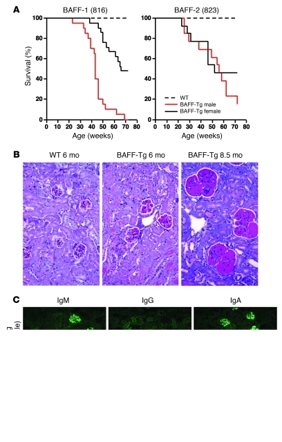
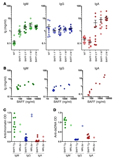
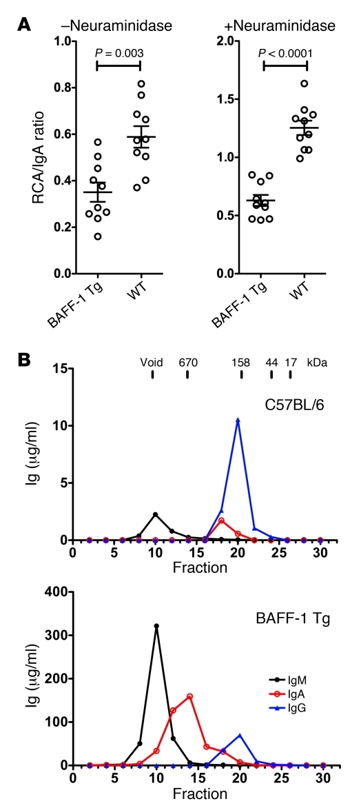
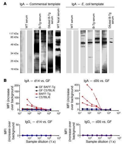
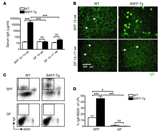
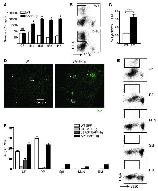
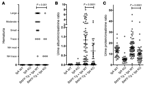
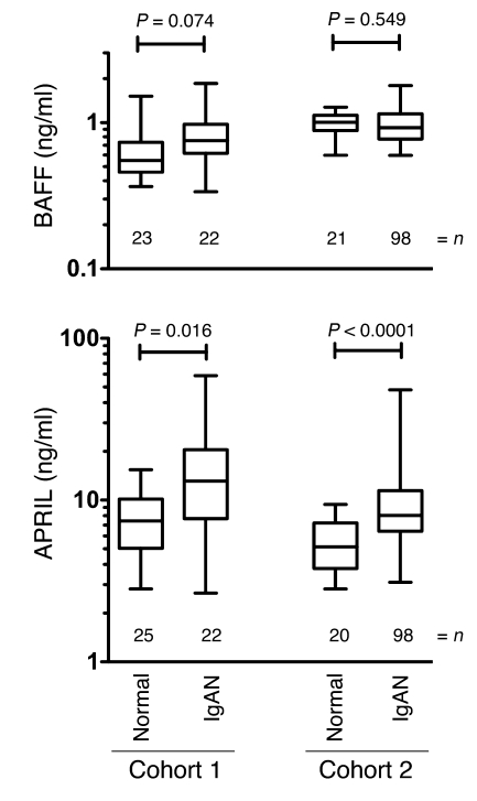
B cell activation factor of the TNF family (BAFF) is a potent B cell survival factor. BAFF overexpressing transgenic mice (BAFF-Tg mice) exhibit features of autoimmune disease, including B cell hyperplasia and hypergammaglobulinemia, and develop fatal nephritis with age. However, basal serum IgA levels are also elevated, suggesting that the pathology in these mice may be more complex than initially appreciated. Consistent with this, we demonstrate here that BAFF-Tg mice have mesangial deposits of IgA along with high circulating levels of polymeric IgA that is aberrantly glycosylated. Renal disease in BAFF-Tg mice was associated with IgA, because serum IgA was highly elevated in nephritic mice and BAFF-Tg mice with genetic deletion of IgA exhibited less renal pathology. The presence of commensal flora was essential for the elevated serum IgA phenotype, and, unexpectedly, commensal bacteria-reactive IgA antibodies were found in the blood. These data illustrate how excess B cell survival signaling perturbs the normal balance with the microbiota, leading to a breach in the normal mucosal-peripheral compartmentalization. Such breaches may predispose the nonmucosal system to certain immune diseases. Indeed, we found that a subset of patients with IgA nephropathy had elevated serum levels of a proliferation inducing ligand (APRIL), a cytokine related to BAFF. These parallels between BAFF-Tg mice and human IgA nephropathy may provide a new framework to explore connections between mucosal environments and renal pathology.


Figures

Figure 1
Figure 1. Gender-biased development of fatal nephropathy associated with dominant IgA and IgM glomerular deposits in the BAFF-Tg mice.
(A) Cohorts of male and female BAFF-Tg mice and WT controls (n = 13–20 each) were observed for up to 21 months. Cohorts of BAFF-Tg mice for the survival experiments were derived from 2 homozygous founders called BAFF-1 (original 816 line) and BAFF-2 (original 823 line) (35), which had been rederived after receipt from the Garvan Institute. Genome scanning showed that both lines retained some DBA2 from the original transgenic derivation using DBA2 × B6 F1 mice (BAFF-1, 10%; BAFF-2, 18%). The longitudinal study shown here was conducted from 2003 to 2005. This study was repeated from 2006 to 2008, and although onset of severe nephropathy was roughly equivalent (30–40 weeks) and the gender bias was present, we observed enhanced survival at the experiment end compared with that of the 2003 to 2005 cohort (data not shown). The improved survival was also recapitulated in BAFF-1 homozygous mice that retained only 4% DBA2 (see further discussion of this topic in the Methods). (B) Representative WT (6 months) and BAFF-2 Tg kidney tissues reveal increased mesangial matrix in BAFF-Tg mice (PAS stain; original magnification, ×200). (C) Representative IgA, IgG, and IgM deposits in BAFF-1 Tg mice versus WT mice (original magnification, ×200).
Figure 2
Figure 2. Dramatically elevated serum IgA in BAFF-Tg mice correlating with serum BAFF levels.
Autoreactivity is limited to the IgM isotype. (A) Serum IgM, IgG, and IgA levels in a cohort of BAFF-1 Tg mice and BAFF-2 Tg mice at 3 to 4 months of age compared with those of matched WT controls. (B) Serum levels of IgA, but not IgM or IgG, are correlated with serum BAFF levels, as measured by ELISA in the BAFF-2 female cohort in A. (C) Antinuclear autoreactivity is observed only in the IgM isotype in BAFF-1 Tg mice. Antichromatin and (D) anti-dsDNA antibodies were evaluated in male BAFF-1 Tg mice and C57BL/6 mice (both 6 months old). Open circles show serum autoantibody levels from a diseased 3-month-old MRL/lpr mouse for reference. Individual symbols represent individual mice; horizontal bars represent the mean ± SEM. F, female; M, male.
Figure 3
Figure 3. Accumulation of underglycosylated IgA and polymeric IgA in the serum of BAFF-Tg mice.
(A) After IgA capture with an isotype-specific antibody, samples were or were not pretreated with neuraminidase, and the glycosylation state was then probed with RCA-1 lectin to detect terminal galactose residues. Relative levels of glycosylation are expressed as a binding ratio (OD lectin detection/OD anti-Ig detection). For reference, a mouse myeloma monoclonal IgA that had low levels of terminal galactose gave ratios of 0.1 and 0.25 (– and + neuraminidase, respectively). Individual symbols represent individual mice; horizontal bars represent the mean ± SEM. (B) Size-exclusion chromatography fractionation of IgA in sera of male C57BL/6 and male BAFF-1-Tg mice, showing the highly polymeric nature of the IgA and increased concentration of IgM in the BAFF-Tg mice.
Figure 4
Figure 4. Evidence for commensal specificity of serum IgA from BAFF-Tg mice.
(A) Lysed commensal bacteria or laboratory strain E. coli were used as the target antigens in a Western blot, using serum or fecal supernatant from WT or BAFF-2 Tg (B-Tg) mice as primary antibody and anti–IgA-HRP as secondary antibody. Blots shown are representative of 3 independent experiments. (B) Serum IgA (top) and IgG levels (bottom) from ASF-reconstituted GF WT mice versus BAFF-2 Tg mice were examined by flow cytometry for specific surface binding to intact Lacto­bacillus murinus, a verified bacterial species retrieved from the intestinal wash of ASF-colonized mice.
Figure 5
Figure 5. Commensal bacteria are necessary for hyper-IgA syndrome in BAFF-Tg mice.
(A) Serum IgA measured from SPF and GF C57BL/6 (WT) and BAFF-2 Tg mice measured by ELISA. At least 4 mice were analyzed per group (***P < 0.001). (B) Immunofluorescence staining of frozen sections of kidneys from SPF and GF C57BL/6 and BAFF-2 Tg mice stained for IgA (green). Images are representative of analysis of at least 8 mice per group (original magnification, ×100). Arrows identify glomeruli that do not exhibit IgA deposition. (C) Representative FACS profiles of IgA+ PCs in the gut LP of SPF versus GF C57BL/6 and BAFF-2 Tg mice. (D) Quantification of IgA+ PCs from C. Analysis was performed on 12-week-old BAFF-2 Tg mice versus C57BL/6 mice. Note that BAFF levels in GF BAFF-2 Tg mice were approximately equal to those observed for SPF BAFF-Tg mice (1,733.2 ± 660.8 pg/ml; n =12). LPL, LP lymphocyte. Bars in A and D represent the mean ± SEM. *P < 0.05, ***P < 0.001.
Figure 6
Figure 6. Hyper-IgA phenotype in BAFF-Tg mice is restored upon recolonization with commensal flora.
(A) Average serum IgA levels in GF C57BL/6 (n = 5) and BAFF-2 Tg (n = 7) mice after introduction of ASF at 6 weeks of age as measured by ELISA. *P < 0.05. (B) Representative FACS and (C) frequency of IgA+ PCs isolated from small intestinal LP of C57BL/6 and BAFF-2 Tg mice after 45 days of recolonization. ***P < 0.01. (D) Representative images of IgA immunofluorescence staining of frozen sections of kidneys from C57BL/6 and BAFF-2 Tg mice recolonized for 45 days (original magnification, ×200). Arrows indicate individual glomeruli. (E) Representative FACS analysis and (F) quantification of IgA+ PCs in different anatomical sites after commensal recolonization contrasted with SPF BAFF-2 Tg mice. A GF reconstitution experiment was performed twice with similar results on 8-week-old BAFF-2 Tg mice versus age-matched C57BL/6 mice. Spl, spleen. Bars in A, C, and F represent the mean ± SEM.
Figure 7
Figure 7. Loss of IgA restores kidney function in BAFF-Tg mice.
Male BAFF-1 Tg mice bred onto a WT or IgA-deficient background were assessed at 13.5 to 15 months of age for the renal damage parameters: (A) hematuria, (B) urinary ratio of microalbumin/creatinine, and (C) urinary ratio of total protein/creatinine. 36 IgA+/+, 28 IgA–/–, 72 BAFF-Tg × IgA+/+, and 48 BAFF-Tg × IgA–/– mice were used, except for hematuria assessment, for which 21 IgA+/+, 23 IgA–/–, 50 BAFF-Tg × IgA+/+, and 33 BAFF-Tg × IgA–/– animals were analyzed ± SEM. Individual symbols represent individual mice; horizontal bars represent the mean ± SEM.
Figure 8
Figure 8. Elevated APRIL and less affected BAFF levels in the serum of patients with IgAN.
Sera from 2 separate groups of biopsy-confirmed patients with IgAN, cohort 1 (Toronto) and cohort 2 (Alabama), were subjected to ELISA quantification of BAFF and APRIL. Box and whisker plots show the 5%–95% limits of the range, and significance was determined by the Mann-Whitney test. The numbers represent the number of patients.

Similar articles

Cited by

References

    1. Fagarasan S, Kawamoto S, Kanagawa O, Suzuki K. Adaptive immune regulation in the gut: T cell-dependent and T cell-independent IgA synthesis. Annu Rev Immunol. 2010;28:243–273. doi: 10.1146/annurev-immunol-030409-101314. - DOI - PubMed
    1. Macpherson AJ, McCoy KD, Johansen FE, Brandtzaeg P. The immune geography of IgA induction and function. Mucosal Immunol. 2008;1(1):11–22. doi: 10.1038/mi.2007.6. - DOI - PubMed
    1. Chervonsky AV. Influence of microbial environment on autoimmunity. Nat Immunol. 2010;11(1):28–35. doi: 10.1038/ni.1801. - DOI - PubMed
    1. Brandtzaeg P. Function of mucosa-associated lymphoid tissue in antibody formation. Immunol Invest. 2010;39(4–5):303–355. doi: 10.3109/08820131003680369. - DOI - PubMed
    1. Yuvaraj S, et al. Evidence for local expansion of IgA plasma cell precursors in human ileum. J Immunol. 2009;183(8):4871–4878. doi: 10.4049/jimmunol.0901315. - DOI - PubMed

Publication types

MeSH terms