Review
. 2011 Dec;115(6):1338-48.
doi: 10.1097/ALN.0b013e3182315d93.

Structural studies of the actions of anesthetic drugs on the γ-aminobutyric acid type A receptor

Affiliations
Review

Structural studies of the actions of anesthetic drugs on the γ-aminobutyric acid type A receptor

Gustav Akk et al. Anesthesiology. 2011 Dec.

Abstract

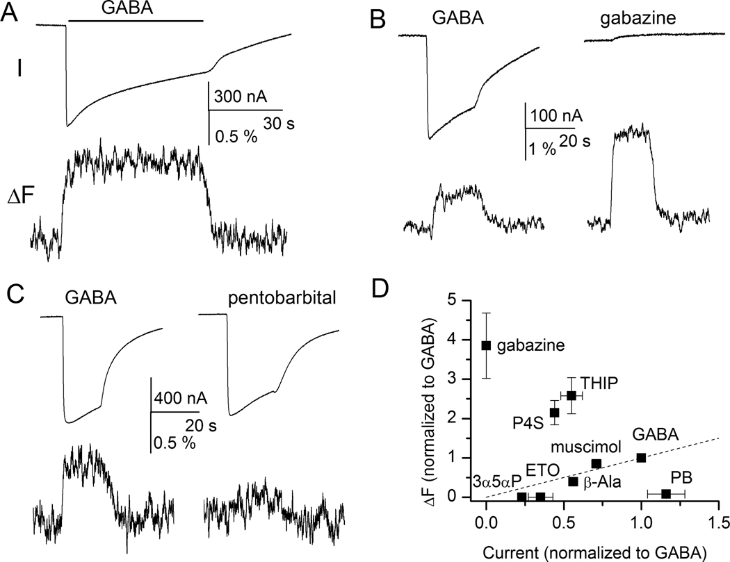
The γ-aminobutyric acid type A receptor is the major transmitter-gated inhibitory channel in the central nervous system. The receptor is a target for anesthetics, anticonvulsants, anxiolytics, and sedatives whose actions facilitate the flow of chloride ions through the channel and enhance the inhibitory tone in the brain. Both the kinetic and structural aspects of the actions of modulators of the γ-aminobutyric acid type A receptor are of great importance to understanding the molecular mechanisms of general anesthesia. In this review, the structural rearrangements that take place in the γ-aminobutyric acid type A receptor during channel activation and modulation are described, focusing on data obtained using voltage-clamp fluorometry. Voltage-clamp fluorometry entails the binding of an environmentally sensitive fluorophore molecule to a site of interest in the receptor, and measurement of changes in the fluorescence signal resulting from activation- or modulation-elicited structural changes. Detailed investigations can provide a map of structural changes that underlie or accompany the functional effects of modulators.


Figures

Figure 1
Figure 1
Organization of the γ-aminobutyric acid type A (GABAA) receptor. (A) A subunit of the GABAA receptor contains a long aminoterminal region, followed by three transmembrane domains (M1–M3), a long cytoplasmic loop, the fourth membrane-spanning segment (M4), and a short carboxyterminal domain. The M2 domain is a major contributor to the central pore. (B) Topology of a subunit showing the region of the subunit contributing to the transmitter binding site (G). The neurosteroid and etomidate binding sites are located in the membrane-spanning domains. A functional receptor is formed of five homologous subunits organized around a central Cl conducting pore. (C) A top view (cross section) of the receptor demonstrating the principal locations of the transmitter binding sites (G) at the β-α subunit interfaces. The β subunit contributes the primary (or, "+") side and the α subunit contributes the complementary (or, "−") side of the transmitter binding site. The receptor contains two pairs of β and α subunits. The fifth subunit (blue in the figure) may be a third α or β subunit, or a γ, δ, or ε subunit. The nature of the subunit in the fifth position can have a strong effect on the functional properties of the receptor. Benzodiazepines interact with a site at the α-γ subunit interface. Some receptors (e.g., ρ subunit-containing) form as homopentamers having the same major structural features.
Figure 2
Figure 2
Voltage-clamp fluorometry. (A) Structures of two commonly-used fluorophores: Alexa 546 maleimide (Alexa 546) and tetramethylrhodamine maleimide (TMRM). For comparison, structure of the transmitter γ-aminobutyric acid (GABA) is shown next to the fluorophores. (B) The maleimide group of the fluorophore binds to the thiol group of the cysteine residue via a maleimide-thiol reaction to form a carbon-sulfur bond. (C) Schematic of the experimental setup. The receptors are expressed in Xenopus oocytes. The oocyte is placed in a custom-made chamber exposing a portion of the membrane through a 0.8 mm pinhole to the lower compartment. The agonist and modulators are applied through the lower compartment. The oocyte is impaled in the top compartment. Two-electrode voltage-clamp is used to record the currents. The chamber is placed into a Petri dish and an inverted microscope, equipped with the appropriate filters, is used to illuminate the oocyte and collect fluorescence.
Figure 3
Figure 3
The extracellular domain of the γ-aminobutyric acid type A (GABAA) receptor portraying the side view at the β2-α1 subunit interface. The β subunit is shown in cyan, the α subunit is shown in pink. The residues which respond with fluorescence change (ΔF) to applications of agonist are shown in blue in the β subunit, and in red in the α subunit. The numbering pertains to the rat α1 and β2 subunits. Sites for which homologous residues in the ρ1 receptor demonstrate ΔF are shown in gray. For those residues, the numbering derives from the human ρ1 sequence. The transmitter binds in the intersubunit cavity near the residues Y241 and L127. A summary of data from fluorescence recordings from these sites is given in table 1. In fluorescence recordings the residues are mutated to cysteines which are then labelled with environmentally-sensitive fluorescent probes. For size comparison, a commonly-used fluorophore, Alexa 546 maleimide, is shown next to the receptor structure.
Figure 4
Figure 4
Sample current and fluorescence recordings from the α1L127Cβ2γ2 receptor labeled with A5m. Receptors were expressed in Xenopus oocutes, and studied with two-electrode voltage clamp . (A) Exposure to γ-aminobutyric acid (GABA) elicits an inward current response (I) and an increase in fluorescence intensity (ΔF) suggesting that the environment around the fluorophore becomes more hydrophobic during channel activation. Channel desensitization does not affect fluorescence changes. (B) Exposure to the competitive antagonist gabazine does not activate the receptor but elicits fluorescence change (ΔF). For comparison, responses to GABA from the same cell are also shown. (C) Activation by GABA but not pentobarbital induces ΔF. Both sets of traces are from the same cell. (D) Relationship between current and fluorescence change. The responses are normalized to those to GABA. The normalized ΔF is plotted as a function of normalized current response. P4S = piperidine-4-sulfonic acid; THIP =4,5,6,7-tetrahydroisoxazolo[5,4-c]pyridin-3-ol; 3α5αP = allopregnanolone; ETO = etomidate; β-Ala = β-alanine, PB = pentobarbital. Panels A, C, and D are reproduced with permission from Akk et al .

References

    1. Bonin RP, Orser BA. GABAA receptor subtypes underlying general anesthesia. Pharmacol Biochem Behav. 2008;90:105–112. - PubMed
    1. Franks NP. General anaesthesia: From molecular targets to neuronal pathways of sleep and arousal. Nat Rev Neurosci. 2008;9:370–386. - PubMed
    1. Jones-Davis DM, Macdonald RL. GABAA receptor function and pharmacology in epilepsy and status epilepticus. Curr Opin Pharmacol. 2003;3:12–18. - PubMed
    1. Rogers CJ, Twyman RE, Macdonald RL. Benzodiazepine and β-carboline regulation of single GABAA receptor channels of mouse spinal neurones in culture. J Physiol. 1994;475:69–82. - PMC - PubMed
    1. Akk G, Bracamontes JR, Covey DF, Evers A, Dao T, Steinbach JH. Neuroactive steroids have multiple actions to potentiate GABAA receptors. J Physiol. 2004;558:59–74. - PMC - PubMed

Publication types