. 2011 Sep 1;2(9):e205.
doi: 10.1038/cddis.2011.91.

Generation and characterization of antibodies specific for caspase-cleaved neo-epitopes: a novel approach

Affiliations

Generation and characterization of antibodies specific for caspase-cleaved neo-epitopes: a novel approach

X Ai et al. Cell Death Dis. .

Abstract

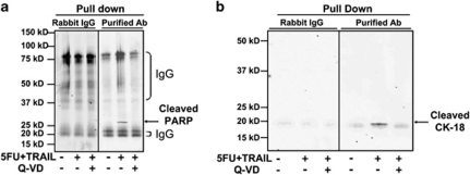
Apoptosis research has been significantly aided by the generation of antibodies against caspase-cleaved peptide neo-epitopes. However, most of these antibodies recognize the N-terminal fragment and are specific for the protein in question. The aim of this project was to create antibodies, which could identify caspase-cleaved proteins without a priori knowledge of the cleavage sites or even the proteins themselves. We hypothesized that many caspase-cleavage products might have a common antigenic shape, given that they must all fit into the same active site of caspases. Rabbits were immunized with the eight most prevalent exposed C-terminal tetrapeptide sequences following caspase cleavage. After purification of the antibodies we demonstrated (1) their specificity for exposed C-terminal (but not internal) peptides, (2) their ability to detect known caspase-cleaved proteins from apoptotic cell lysates or supernatants from apoptotic cell culture and (3) their ability to detect a caspase-cleaved protein whose tetrapeptide sequence differs from the eight tetrapeptides used to generate the antibodies. These antibodies have the potential to identify novel neo-epitopes produced by caspase cleavage and so can be used to identify pathway-specific caspase cleavage events in a specific cell type. Additionally this methodology may be applied to generate antibodies against products of other proteases, which have a well-defined and non-promiscuous cleavage activity.


Figures

Figure 1
Figure 1
Strategy of immunization. (a) In all, 262 caspase-cleavage sites were analyzed to determine the most prevalent C-terminally exposed (after caspase cleavage) tetrapeptide sequences. Based on this analysis, the top eight were chosen. The tetrapeptides were joined to two different eight amino-acids ‘linker'sequences (Scramble 1 and Scramble 2), chosen to closely resemble native rabbit peptides in order to minimize generation of antibodies to these irrelevant parts of the peptide. (b) The cohort of rabbits immunized with the mixture of eight Scramble 1 peptides were boosted once with the Scramble 2 peptides, and vise versa, such that the only commonality between immunization and boost was the C-terminal DXXD sequences. Scramble 3 was utilized during purification to decrease the isolation of irrelevant antibodies to the other linker sequences
Figure 2
Figure 2
Purification of antibodies. (a) Purification scheme. The serum was fractionated with 50% ammonium sulfate and subsequent DEAE column as described in ‘Materials and Methods'. The flow-through from the DEAE column was further purified with eight peptides (Scramble 3-DXXD) conjugated to sulfolink affinity resin. The eluate was collected and the titer of antibodies was tested by ELISA. (b) SDS-PAGE and western blot showed the purification progress of the antibodies. (c) ELISA showed the titer of antibodies purified from the serum of rabbit 2 against eight specific peptides (open circles). Scramble 1-DXXDZZ peptides were used as negative control to show the titer of antibodies against uncleaved substrate sites (filled circles). (d) The high O.D. values of Scramble 3-DEVD-coated wells compared with Scramble 1-, Scramble 2-, Scramble 3-, and OMPC-coated wells are an example of the specificity of the purified antibodies, in all six rabbits tested
Figure 3
Figure 3
Purified antibodies specifically recognize cleaved caspase substrates with a pattern of ‘DXXD'. Apoptosis was induced by 5-FU and TRAIL in HCT116 cells as described in ‘Materials and Methods'. QVD-OPH was added to block caspase activities as indicated. Cell lysate from 107 cells was immuno-precipitated with 20 μg of purified antibodies, then detected with antibodies against PARP (a), caspase 6 (b) or cleaved CK-18 (c), separately. The purified antibodies specifically recognize not only the cleaved PARP (DEVD) and caspase 6 (DVVD) whose caspase cleavage sites are one of the eight peptides used for immunization, but also the cleaved CK-18 (DALD), whose tetrapeptide cleavage site was not one of the eight peptides, but has aspartic acid at the P1 and P4 position. However, the purified antibodies do not recognize the cleaved caspase 7 (IQAD) or lamin A (VEID) whose tetrapeptide cleavage sites has a ‘D' at P1 but not P4 position (data not shown). Rabbit IgGs in the immune precipitate were detected as artifacts in western blots (as labeled) by the secondary antirabbit antibodies. This was due to the fact that both the NEAs and the detection antibodies (i.e. anti-PARP and anti-caspase 6) were raised in the same species (rabbit). Furthermore, to avoid precipitation, samples were not boiled before loading onto gels, therefore IgG oligmers were seen on the blots. (d) Purified antibodies specifically pull down cleaved recombinant CK-18. A total of 10 μg of recombinant CK-18 was cleaved with caspase 3 in vitro. QVD-OPH was added to block caspase activities as indicated. The reaction was precipitated with purified antibodies and then detected with antibody against cleaved CK-18. (e) Peptides with a ‘D' at P1, or P1 and P4 (IETD, LQTD, SECD, DCRD, FRHD and FAED) were coated on the ELISA plates, and probed with purified antibodies. Scramble 3-DETD and Scramble 3 peptides were coated as positive and negative control, respectively. The antibodies only recognize the peptides with aspartic acids at both the P1 and P4 position (i.e. DCRD and DETD), but not the ones with a ‘D' only at P1 position
Figure 4
Figure 4
Purified antibodies can immunoprecipitate different proteins from apoptotic cell lysates compared with lysates of healthy or caspase-inhibited cells. Immunoprecipitated cell lysates from cells as in Figure 3 were also resolved by SDS-PAGE and protein bands were visualized by Coomassie blue staining. Clear differences were observed; arrows indicate for example, at least five (as yet unidentified) bands distinctly more visible in the apoptotic cell lysates compared with lysates from healthy or caspase-inhibited cells. Sections of the gel were cut and subjected to trypsin digestion and mass spectrometry identification. Three known caspase substrates (CK-18, DNA PK and EF1-B) and a novel one (60S ribosomal protein L21) were identified in apoptotic cell lysates
Figure 5
Figure 5
Purified antibodies specifically recognize cleaved PARP and CK-18 in apoptotic media. Apoptosis was induced by 5-FU and TRAIL in HCT116 cells as described in ‘Materials and Methods'. QVD-OPH was added to block caspase activities as indicated. Cell media from 107 cells was concentrated and immuno-precipitated with 20 μg of purified antibodies, then detected with antibodies against PARP (a), or cleaved CK-18 (b), separately. The purified antibodies specifically recognize the cleaved PARP and CK-18 released to the media. The rabbit IgG bands from the antibodies were labeled as well

Similar articles

Cited by

References

    1. Fulda S, Debatin KM. Extrinsic versus intrinsic apoptosis pathways in anticancer chemotherapy. Oncogene. 2006;25:4798–4811. - PubMed
    1. Fulda S. Exploiting apoptosis pathways for the treatment of pediatric cancers. Pediatr Blood Cancer. 2009;53:533–536. - PubMed
    1. Sarker D, Workman P. Pharmacodynamic biomarkers for molecular cancer therapeutics. Adv Cancer Res. 2007;96:213–268. - PubMed
    1. Amur S, Frueh FW, Lesko LJ, Huang SM. Integration and use of biomarkers in drug development, regulation and clinical practice: a US regulatory perspective. Biomark Med. 2008;2:305–311. - PubMed
    1. Ward T, Cummings J, Dean E, Greystoke A, Hou JM, Backen A, et al. Biomarkers of apoptosis. Br J Cancer. 2008;99:841–846. - PMC - PubMed