. 2012 Apr;54(4):369-82.
doi: 10.1007/s00234-011-0948-x. Epub 2011 Sep 1.

Pipeline embolization device (PED) for neurovascular reconstruction: initial experience in the treatment of 101 intracranial aneurysms and dissections

Affiliations

Pipeline embolization device (PED) for neurovascular reconstruction: initial experience in the treatment of 101 intracranial aneurysms and dissections

Sebastian Fischer et al. Neuroradiology. 2012 Apr.

Abstract

Introduction: The purpose of this study was to evaluate the safety and efficacy of the recently available flow diverter "pipeline embolization device" (PED) for the treatment of intracranial aneurysms and dissections.

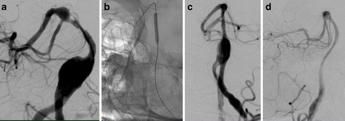
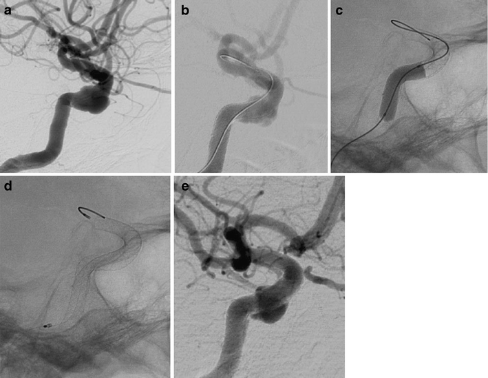
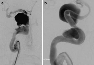
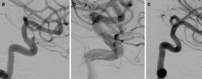
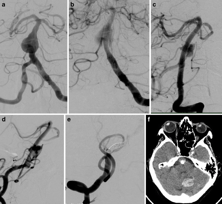
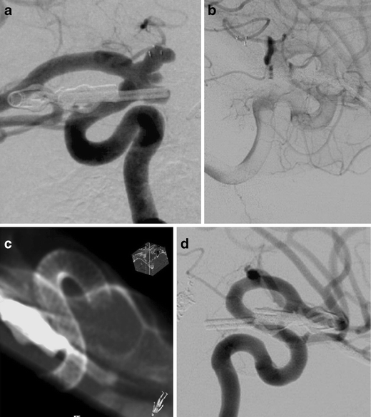
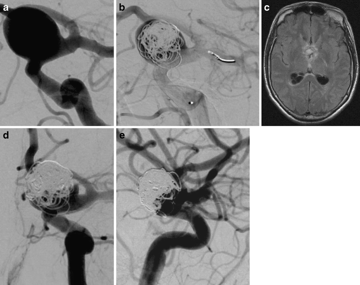
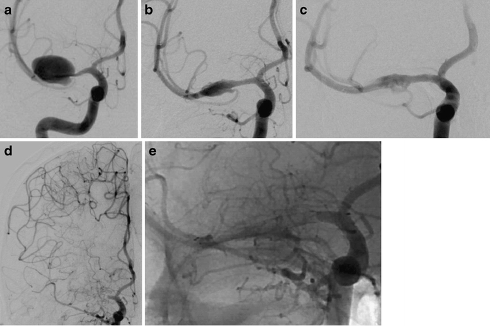
Methods: Eighty-eight consecutive patients underwent an endovascular treatment of 101 intracranial aneurysms or dissections using the PED between September 2009 and January 2011. The targeted vessels include 79 (78%) in the anterior circulation and 22 (22%) in the posterior circulation. We treated 96 aneurysms and 5 vessel dissections. Multiple devices were implanted in 67 lesions (66%).

Results: One technical failure of the procedure was encountered. Immediate exclusion of the target lesion was not observed. Angiographic follow-up examinations were carried out in 80 patients (91%) with 90 lesions and revealed complete cure of the target lesion(s) in 47 (52%), morphological improvement in 32 lesions (36%), and no improvement in 11 lesions (12%). Six major complications were encountered: one fatal aneurysm rupture, one acute and one delayed PED thrombosis, and three hemorrhages in the dependent brain parenchyma.

Conclusion: Our experience reveals that the PED procedure is technically straightforward for the treatment of selected wide-necked saccular aneurysms, fusiform aneurysms, remnants of aneurysms, aneurysms with a high likelihood of failure with conventional endovascular techniques, and dissected vessels. While vessel reconstruction, performed after dissection, is achieved within days, remodeling of aneurysmal dilatations may take several months. Dual platelet inhibition is obligatory. Parenchymal bleeding into brain areas dependent on the target vessel is uncommon.


Figures

Fig. 1
Fig. 1
Fusiform dilatation of the basilar artery with associated stenosis (a). Preparatory balloon angioplasty (b), followed by PED deployment [3 × 4.5 × 20, 4.75 × 20, 4.75 × 18, 5.0 × 20, 3.5 × 20, 3.0 × 20] (c). Angiographic follow-up 4 months later shows remodeling of the dilated vessel (d)
Fig. 2
Fig. 2
Cavernous aneurysm of the right ICA (a). Poor wall apposition after deployment of the second PED [2 × 4.5/20] (b). Adaptation of the PEDs to the vessel wall using a 4 × 15-mm Ascent balloon (c) results in secondary expansion of the second device with close contact between its outer surface and the vessel wall (d). Angiographic follow-up 3 months later shows a decrease of the aneurysm size and a regular reconstruction of the parent artery (e)
Fig. 3
Fig. 3
Deployment of 19 PEDs in a patient with large aneurysms of the right ICA, presenting with diplopia and severe headache. Injection of the right ICA after direct puncture shows fusiform aneurysms of the petrous and cavernous segments separated by a relative stenosis (a). Reconstruction of the vessel with 19 PEDs after preparatory balloon angioplasty (b). MRI 3 days later shows thrombosis of both aneurysms. In the meantime, the patient has improved clinically
Fig. 4
Fig. 4
Progressive vessel remodeling after flow diversion. Injection of the left ICA with a paraophthalmic aneurysm (a). Deployment of three PEDs induced contrast stagnation in the aneurysm (b). After 4 months, complete obliteration of the aneurysm was confirmed (c)
Fig. 5
Fig. 5
Large fusiform aneurysm of the basilar trunk with brainstem compression (a). Contrast stagnation within the aneurysm after PED deployment [2 × 3/18 mm] (b). Shrinkage of the aneurysm 8 months later with preserved AICAs (c). Reperfusion and recurrent growth of the fusiform aneurysm 11 months after the treatment (d) induced by flow coming from the right VA. Coil occlusion of the right V4 segment (e). Cerebellar hemorrhage 1 day after coil occlusion treated conservatively (f)
Fig. 6
Fig. 6
Four de novo aneurysms of the right paraclinoid and supraclinoid ICA, following microsurgical clipping of an aneurysm in the same location years ago (a). Stasis of contrast material in the aneurysms after deployment of two PEDs [2 × 2.5/20 mm] (b). DynaCT confirmed the full expansion of the PEDs (c). Obliteration of the four aneurysms with reconstruction of the lumen of the right ICA was confirmed at the first follow-up DSA after 4 months (d)
Fig. 7
Fig. 7
Rupture of a wide-necked paraclinoid aneurysm 3 days after loose coiling and deployment of three PEDs [4/14, 4.25/20, 4.25/14]. Aneurysm prior to treatment (a) and immediately after coiling and PED implantation with stagnating contrast medium (b). Four days later, the patient presented severe headache and visual loss. MRI showed a massive intraventricular hemorrhage (c). DSA confirmed a neck remnant of the aneurysm with thrombosis of the fundus 3 h later (d). Late-phase DSA images revealed an accumulation of contrast medium outside the contour of the aneurysm sac adjacent to the aneurysm dome (e)
Fig. 8
Fig. 8
Acute thrombosis of a PED deployed within two previously implanted self-expanding stents. Dissecting, partially thrombosed aneurysm of the right M1 segment (a). An Enterprise and a Solitaire stent were coaxially deployed to enforce the vessel wall and to redirect blood flow in June 2009. Follow-up DSA showed further but incomplete thrombosis of the aneurysm (b). Two PEDs [2.5 × 20, 2.5 × 18] were deployed within the two stents (c). Despite dual antiplatelet antiaggregation, acute thrombosis of PED and stents occurred 5 days later with significant MCA ischemia but without a neurological deficit (d, e)

References

    1. Henkes H, Bose A, Felber S, Miloslavski E, Berg-Dammer E, Kühne D. Endovascular coil occlusion of intracranial aneurysms assisted by a novel self-expandable nitinol microstent (Neuroform) Interv Neuroradiol. 2002;30:107–119. - PMC - PubMed
    1. Shapiro M, Babb J, Becske T, Nelson PK. Safety and efficacy of adjunctive balloon remodeling during endovascular treatment of intracranial aneurysms: a literature review. AJNR Am J Neuroradiol. 2008;29:1777–1781. doi: 10.3174/ajnr.A1216. - DOI - PMC - PubMed
    1. Augsburger L, Farhat M, Reymond P, Fonck E, Kulcsar Z, Stergiopulos N, Rüfenacht DA. Effect of flow diverter porosity on intraaneurysmal blood flow. Clin Neuroradiol. 2009;19:204–214. doi: 10.1007/s00062-009-9005-0. - DOI - PubMed
    1. Henkes H, Reinartz J, Preiss H, Miloslavski E, Kirsch M, Kühne D. Endovascular treatment of small intracranial aneurysms: three alternatives to coil occlusion. Minim Invasive Neurosurg. 2006;49:65–69. doi: 10.1055/s-2005-919150. - DOI - PubMed
    1. Felber S, Henkes H, Weber W, Miloslavski E, Brew S, Kühne D. Treatment of extracranial and intracranial aneurysms and arteriovenous fistulae using stent grafts. Neurosurgery. 2004;55:631–638. doi: 10.1227/01.NEU.0000134455.02947.1F. - DOI - PubMed