

Development and characterization of an organotypic model of Barrett's esophagus
- PMID: 21882191
- PMCID: PMC3352665
- DOI: 10.1002/jcp.23007
Development and characterization of an organotypic model of Barrett's esophagus
Abstract
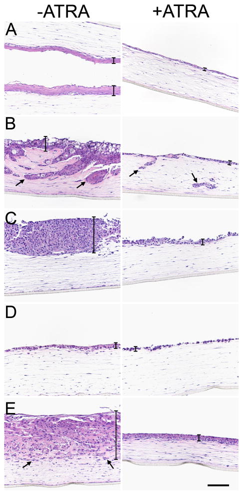
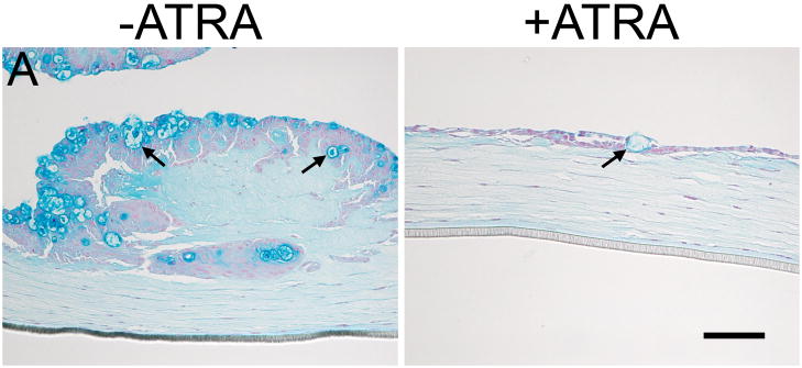
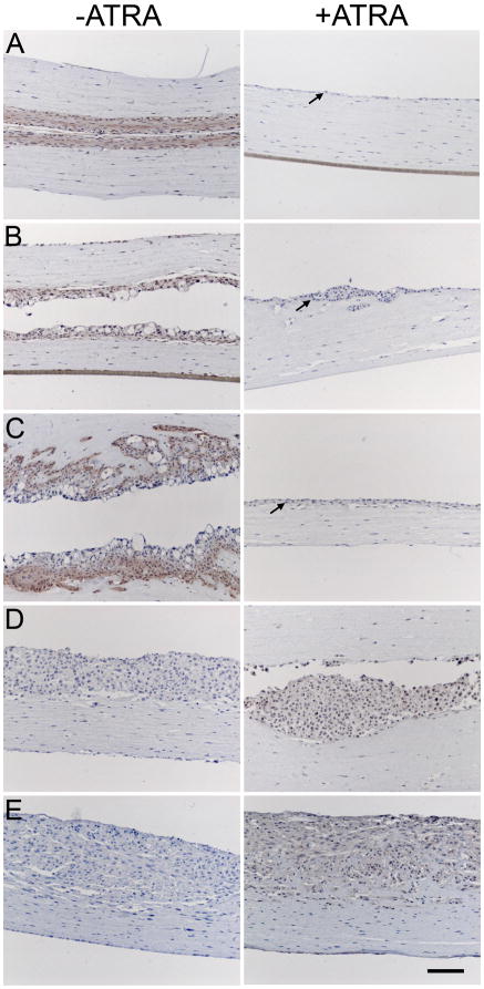
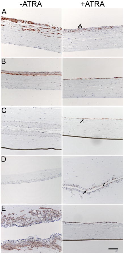
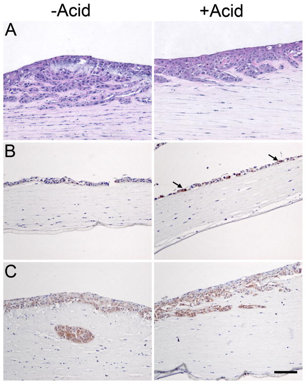
Understanding the molecular and cellular processes underlying the development, maintenance, and progression of Barrett's esophagus (BE) presents an empirical challenge because there are no simple animal models and standard 2D cell culture can distort cellular processes. Here we describe a three-dimensional (3D) cell culture system to study BE. BE cell lines (CP-A, CP-B, CP-C, and CP-D) and esophageal squamous keratinocytes (EPC2) were cultured on a matrix consisting of esophageal fibroblasts and collagen. Comparison of growth and cytokeratin expression in the presence of all-trans retinoic acid or hydrochloric acid was made by immunohistochemistry and Alcian Blue staining to determine which treatments produced a BE phenotype of columnar cytokeratin expression in 3D culture. All-trans retinoic acid differentially affected the growth of BE cell lines in 3D culture. Notably, the non-dyplastic metaplasia-derived cell line (CP-A) expressed reduced squamous cytokeratins and enhanced columnar cytokeratins upon ATRA treatment. ATRA altered the EPC2 squamous cytokeratin profile towards a more columnar expression pattern. Cell lines derived from patients with high-grade dysplasia already expressed columnar cytokeratins and therefore did not show a systematic shift toward a more columnar phenotype with ATRA treatment. ATRA treatment, however, did reduce the squamoid-like multilayer stratification observed in all cell lines. As the first study to demonstrate long-term 3D growth of BE cell lines, we have determined that BE cells can be cultured for at least 3 weeks on a fibroblast/collagen matrix and that the use of ATRA causes a general reduction in squamous-like multilayered growth and an increase in columnar phenotype with the specific effects cell-line dependent.
Copyright © 2011 Wiley Periodicals, Inc.
Figures
Similar articles
-
Aberrant epithelial-mesenchymal Hedgehog signaling characterizes Barrett's metaplasia.Gastroenterology. 2010 May;138(5):1810-22. doi: 10.1053/j.gastro.2010.01.048. Epub 2010 Feb 4. Gastroenterology. 2010. PMID: 20138038 Free PMC article.
-
Barrett's metaplasia develops from cellular reprograming of esophageal squamous epithelium due to gastroesophageal reflux.Am J Physiol Gastrointest Liver Physiol. 2017 Jun 1;312(6):G615-G622. doi: 10.1152/ajpgi.00268.2016. Epub 2017 Mar 23. Am J Physiol Gastrointest Liver Physiol. 2017. PMID: 28336546
-
Inhibition of Notch signaling enhances transdifferentiation of the esophageal squamous epithelium towards a Barrett's-like metaplasia via KLF4.Cell Cycle. 2014;13(24):3857-66. doi: 10.4161/15384101.2014.972875. Cell Cycle. 2014. PMID: 25558829 Free PMC article.
-
Origins of Metaplasia in Barrett's Esophagus: Is this an Esophageal Stem or Progenitor Cell Disease?Dig Dis Sci. 2018 Aug;63(8):2005-2012. doi: 10.1007/s10620-018-5069-5. Dig Dis Sci. 2018. PMID: 29675663 Free PMC article. Review.
-
Modelling Barrett's oesophagus.Biochem Soc Trans. 2010 Apr;38(2):321-6. doi: 10.1042/BST0380321. Biochem Soc Trans. 2010. PMID: 20298176 Review.
Cited by
-
Discovery and validation of Barrett's esophagus microRNA transcriptome by next generation sequencing.PLoS One. 2013;8(1):e54240. doi: 10.1371/journal.pone.0054240. Epub 2013 Jan 23. PLoS One. 2013. PMID: 23372692 Free PMC article.
-
The Microenvironment in Barrett's Esophagus Tissue Is Characterized by High FOXP3 and RALDH2 Levels.Front Immunol. 2018 Jun 18;9:1375. doi: 10.3389/fimmu.2018.01375. eCollection 2018. Front Immunol. 2018. PMID: 29967615 Free PMC article.
-
Modeling human gastrointestinal inflammatory diseases using microphysiological culture systems.Exp Biol Med (Maywood). 2014 Sep;239(9):1108-23. doi: 10.1177/1535370214529388. Epub 2014 Apr 29. Exp Biol Med (Maywood). 2014. PMID: 24781339 Free PMC article. Review.
-
Review: Experimental models for Barrett's esophagus and esophageal adenocarcinoma.Am J Physiol Gastrointest Liver Physiol. 2012 Jun 1;302(11):G1231-43. doi: 10.1152/ajpgi.00509.2011. Epub 2012 Mar 15. Am J Physiol Gastrointest Liver Physiol. 2012. PMID: 22421618 Free PMC article. Review.
-
Esophageal adenocarcinoma models: a closer look.Front Mol Biosci. 2024 Nov 12;11:1440670. doi: 10.3389/fmolb.2024.1440670. eCollection 2024. Front Mol Biosci. 2024. PMID: 39600303 Free PMC article. Review.
References
-
- Aydelotte MB. The Effects of Vitamin A and Citral on Epithelial Differentiation in vitro 2. The Chick Oesophageal and Corneal Epithelia and Epidermis. Journal of Embryology and Experimental Morphology. 1963;11(3):621–635. - PubMed
-
- Bajpai M, Liu J, Geng X, Souza RF, Amenta PS, Das KM. Repeated exposure to acid and bile selectively induces colonic phenotype expression in a heterogeneous Barrett’s epithelial cell line. Lab Invest. 2008;88(6):643–651. - PubMed
-
- Biddle D, Spandau D. Expression of vimentin in cultured human keratinocytes is associated with cell—extracellular matrix junctions. Archives of Dermatological Research. 1996;288(10):621–624. - PubMed
-
- Boch JA, Shields HM, Antonioli DA, Zwas F, Sawhney RA, Trier JS. Distribution of cytokeratin markers in Barrett’s specialized columnar epithelium. Gastroenterology. 1997;112(3):760–765. - PubMed
Publication types
MeSH terms
Substances
Grants and funding
LinkOut - more resources
Full Text Sources
Research Materials
Miscellaneous
