Comparative Study
. 2011 Nov;119(3):594-603.
doi: 10.1111/j.1471-4159.2011.07456.x. Epub 2011 Sep 21.

Diabetic neuropathy enhances voltage-activated Ca2+ channel activity and its control by M4 muscarinic receptors in primary sensory neurons

Affiliations
Comparative Study

Diabetic neuropathy enhances voltage-activated Ca2+ channel activity and its control by M4 muscarinic receptors in primary sensory neurons

Xue-Hong Cao et al. J Neurochem. 2011 Nov.

Abstract

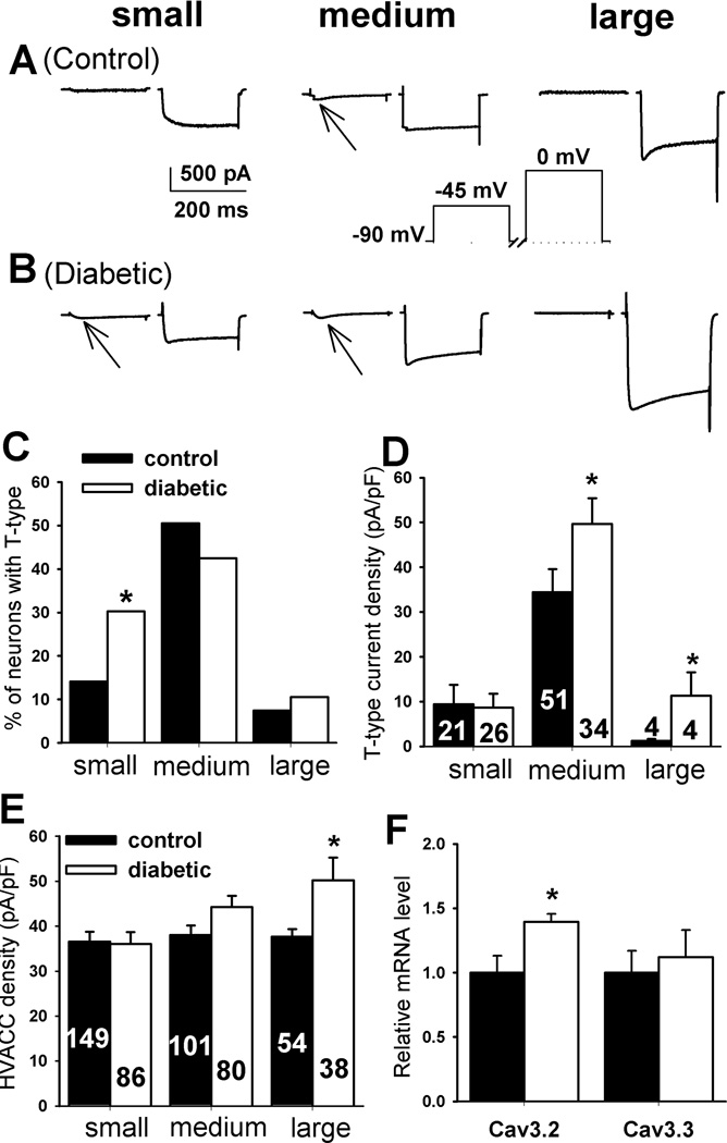
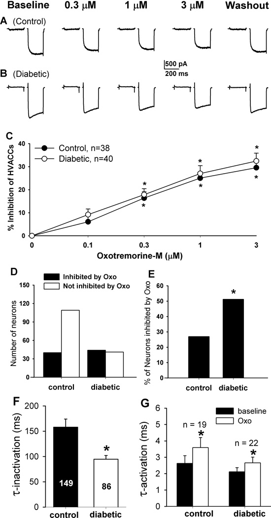
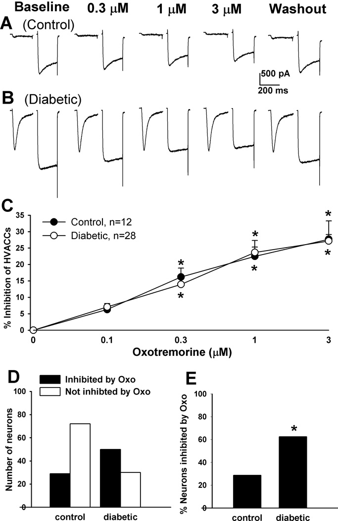
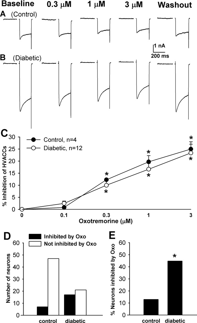
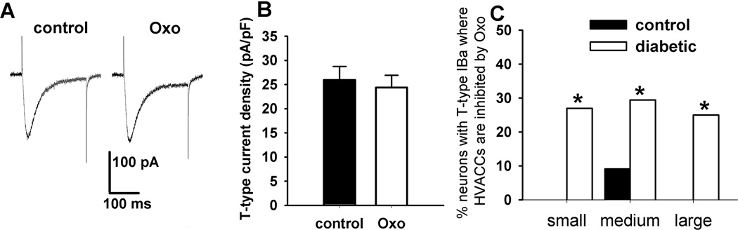
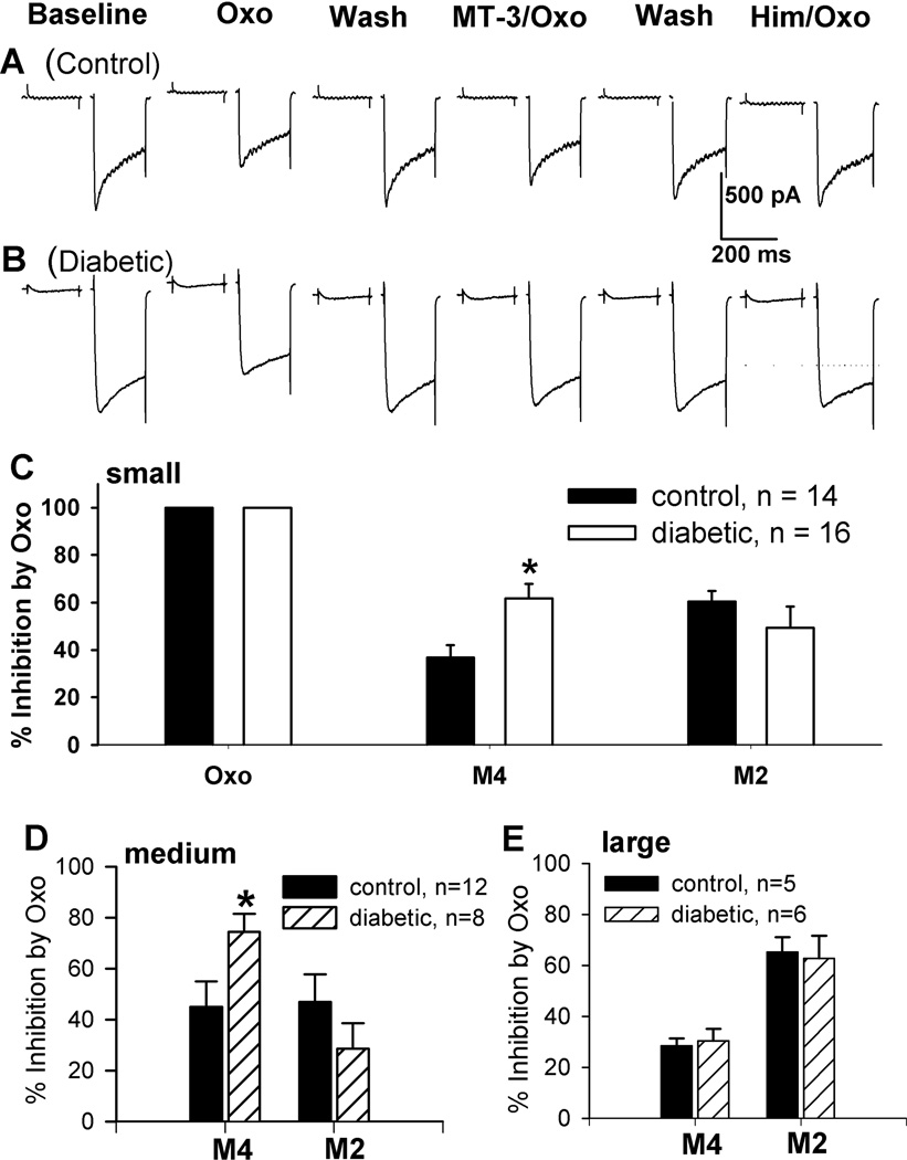
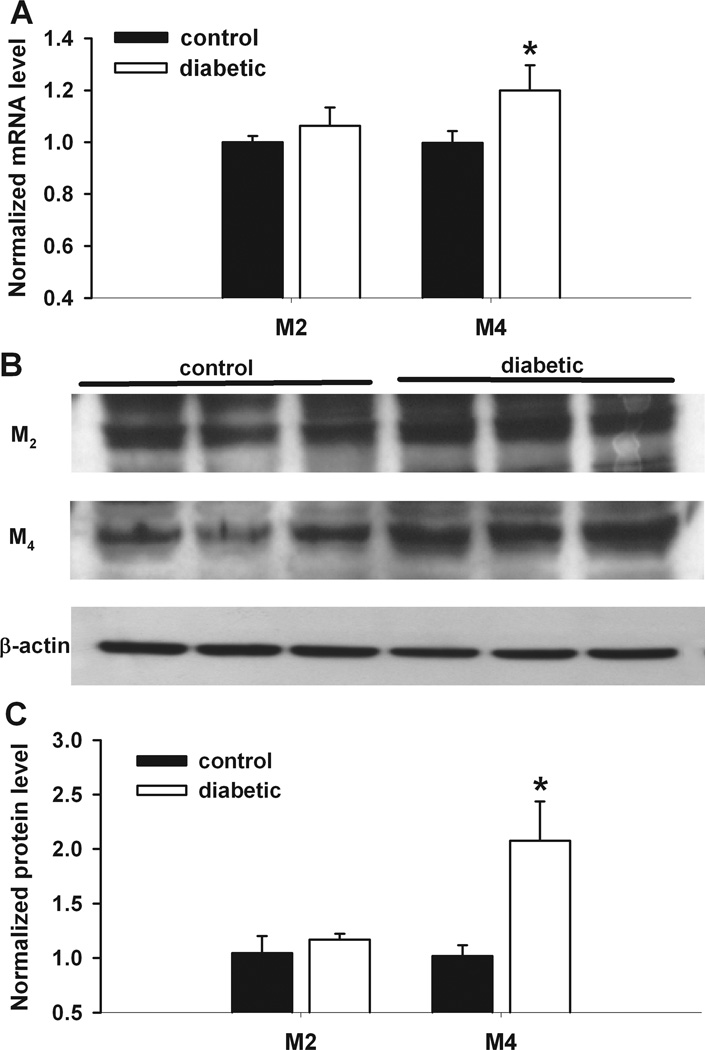
Painful neuropathy is one of the most serious complications of diabetes and remains difficult to treat. The muscarinic acetylcholine receptor (mAChR) agonists have a profound analgesic effect on painful diabetic neuropathy. Here we determined changes in T-type and high voltage-activated Ca(2+) channels (HVACCs) and their regulation by mAChRs in dorsal root ganglion (DRG) neurons in a rat model of diabetic neuropathy. The HVACC currents in large neurons, T-type currents in medium and large neurons, the percentage of small DRG neurons with T-type currents, and the Cav3.2 mRNA level were significantly increased in diabetic rats compared with those in control rats. The mAChR agonist oxotremorine-M significantly inhibited HVACCs in a greater proportion of DRG neurons with and without T-type currents in diabetic than in control rats. In contrast, oxotremorine-M had no effect on HVACCs in small and large neurons with T-type currents and in most medium neurons with T-type currents from control rats. The M(2) and M(4) antagonist himbacine abolished the effect of oxotremorine-M on HVACCs in both groups. The selective M(4) antagonist muscarinic toxin-3 caused a greater attenuation of the effect of oxotremorine-M on HVACCs in small and medium DRG neurons in diabetic than in control rats. Additionally, the mRNA and protein levels of M(4), but not M(2), in the DRG were significantly greater in diabetic than in control rats. Our findings suggest that diabetic neuropathy potentiates the activity of T-type and HVACCs in primary sensory neurons. M(4) mAChRs are up-regulated in DRG neurons and probably account for increased muscarinic analgesic effects in diabetic neuropathic pain.


Conflict of interest statement

The authors declare that they have no conflict of interest regarding the work presented here.

Figures

Figure 1
Figure 1
Changes in the activity of T-type VACCs and HVACCs in DRG neurons of diabetic rats. A, B: typical traces show T-type (indicated by arrows) and HVACC IBa in small, medium, and large DRG neurons from control and diabetic rats. C: summary data show the percentage of small, medium, and large DRG neurons with T-type IBa in control and diabetic rats. D: group data show the current density of T-type IBa of control and diabetic rats. E: summary data show the current density of HVACCs of DRG neurons in control and diabetic rats. F: group data show changes in the mRNA level of Cav3.2 and Cav3.3 subtypes in the DRG from control and diabetic rats (n= 6 samples in each group). Data were presented as mean ± SEM. *P < 0.05, compared with the control group.
Figure 2
Figure 2
Effects of Oxo-M on HVACC IBa in small DRG neurons from control and diabetic rats. A, B: original traces show that Oxo-M dose-dependently inhibited HVACC IBa in small DRG neurons from control and diabetic rats. C: summary data show the dose-response effect of Oxo-M on HVACC IBa in small DRG neurons from control and diabetic group. *P < 0.05, compared with the baseline before Oxo-M application. D: group data show the number of Oxo-M-responsive and Oxo-M-unresponsive small DRG neurons in control and diabetic rats. E: comparison of the percentage of Oxo-M-responsive small DRG neurons in control and diabetic rats. F: the inactivation time constant of HVACC currents in small DRG neurons in diabetic and control rats. G: effects of 1 µM Oxo-M on the activation time constant of HVACCs in small DRG neurons in control and diabetic rats. *P < 0.05, compared with the control or baseline group.
Figure 3
Figure 3
Effects of Oxo-M on HVACC IBa in medium DRG neurons from control and diabetic rats. A, B: representative traces show that Oxo-M dose-dependently inhibited HVACC IBa in medium DRG neurons from control and diabetic rats. C: summary data show the dose-response effect of Oxo-M on HVACC IBa in medium DRG neurons from control and diabetic group. *P < 0.05, compared with the baseline before Oxo-M application. D: group data show the number of Oxo-M-responsive and Oxo-M-unresponsive medium DRG neurons in control and diabetic rats. E: comparison of the percentage of Oxo-M-responsive medium DRG neurons in control and diabetic rats. *P < 0.05, compared with the control group.
Figure 4
Figure 4
Effects of Oxo-M on HVACC IBa in large DRG neurons from control and diabetic rats. A, B: original traces show that Oxo-M dose-dependently inhibited HVACC IBa in large DRG neurons from control and diabetic rats. C: summary data show the dose-response effect of Oxo-M on HVACC IBa in large DRG neurons from control and diabetic group. *P < 0.05, compared with the baseline before Oxo-M application. D: group data show the number of Oxo-M-responsive and Oxo-M-unresponsive large DRG neurons in control and diabetic rats. E: comparison of the percentage of Oxo-M-responsive large DRG neurons in control and diabetic rats. *P < 0.05, compared with the control group.
Figure 5
Figure 5
Lack of effects of Oxo-M on T-type IBa in DRG neurons from control and diabetic rats. A: original traces show that 1 µM Oxo-M had no effect on T-type IBa in a medium DRG neuron from a diabetic rat. B: group data show that Oxo-M had no effect on the current density of T-type IBa in a total of 140 DRG neurons from control and diabetic rats. C: summary data show the percentage of neurons with T-type IBa inhibited by 1 µM Oxo-M in control and diabetic rats. *P < 0.05, compared with the control group.
Figure 6
Figure 6
Contribution of M2 and M4 subtypes to inhibition of HVACC IBa by Oxo-M in DRG neurons from control and diabetic rats. A, B: representative traces show the effect of 1 µM Oxo-M on HVACC IBa in the presence of 50 nM MT-3 or 2 µM himbacine in small DRG neurons from a control and a diabetic rat. C: comparison of the contribution of M2 and M4 subtypes to inhibition on HVACC IBa by Oxo-M in small DRG neurons from control and diabetic rats. D: summary data show M2- and M4-mediated inhibition of HVACC IBa by Oxo-M in medium DRG neurons from control and diabetic rats. E: group data show M2- and M4-mediated inhibition of HVACC IBa by Oxo-M in large DRG neurons from control and diabetic rats. *P < 0.05, compared with the control group.
Figure 7
Figure 7
Changes in the mRNA and protein levels of M2 and M4 subtypes in the DRG of diabetic rats. A: group data show the difference in the mRNA level of M2 and M4 subtypes in the DRG between control and diabetic rats (n = 6 samples in each group). B: original gel images show the protein level of M2 and M4 subunits in the DRG from control and diabetic rats. C: summary data show the M2 and M4 protein levels in the DRG from control and diabetic rats (n = 6 samples in each group). *P < 0.05, compared with the control group.

References

    1. Bell TJ, Thaler C, Castiglioni AJ, Helton TD, Lipscombe D. Cell-specific alternative splicing increases calcium channel current density in the pain pathway. Neuron. 2004;41:127–138. - PubMed
    1. Bernardini N, Levey AI, Augusti-Tocco G. Rat dorsal root ganglia express m1-m4 muscarinic receptor proteins. J Peripher Nerv Syst. 1999;4:222–232. - PubMed
    1. Bourinet E, Alloui A, Monteil A, Barrere C, Couette B, Poirot O, Pages A, McRory J, Snutch TP, Eschalier A, Nargeot J. Silencing of the Cav3.2 T-type calcium channel gene in sensory neurons demonstrates its major role in nociception. Embo J. 2005;24:315–324. - PMC - PubMed
    1. Cai YQ, Chen SR, Han HD, Sood AK, Lopez-Berestein G, Pan HL. Role of M2, M3, and M4 muscarinic receptor subtypes in the spinal cholinergic control of nociception revealed using siRNA in rats. J Neurochem. 2009;111:1000–1010. - PMC - PubMed
    1. Cao XH, Byun HS, Chen SR, Cai YQ, Pan HL. Reduction in voltage-gated K+ channel activity in primary sensory neurons in painful diabetic neuropathy: role of brain-derived neurotrophic factor. J Neurochem. 2010;114:1460–1475. - PMC - PubMed

Publication types

MeSH terms