

Computationally predicted IgE epitopes of walnut allergens contribute to cross-reactivity with peanuts
- PMID: 21883278
- PMCID: PMC3203311
- DOI: 10.1111/j.1398-9995.2011.02692.x
Computationally predicted IgE epitopes of walnut allergens contribute to cross-reactivity with peanuts
Abstract
Background: Cross-reactivity between peanuts and tree nuts implies that similar immunoglobulin E (IgE) epitopes are present in their proteins.
Objective: To determine whether walnut sequences similar to known peanut IgE-binding sequences, according to the property distance (PD) scale implemented in the Structural Database of Allergenic Proteins, react with IgE from sera of patients with allergy to walnut and/or peanut.
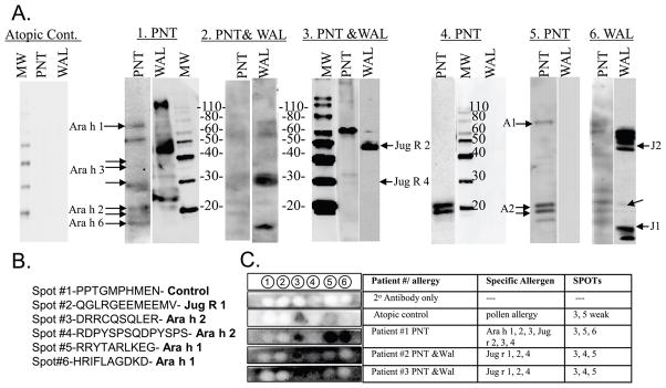
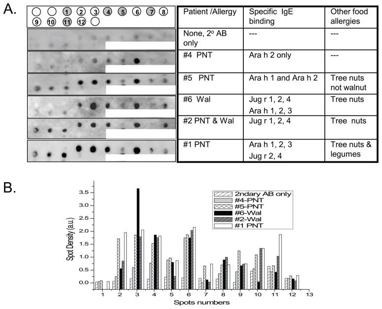
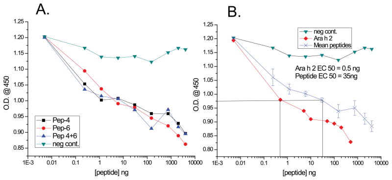
Methods: Patient sera were characterized by western blotting for IgE binding to nut protein extracts and to peptides from walnut and peanut allergens, similar to known peanut epitopes as defined by low PD values, synthesized on membranes. Competitive enzyme-linked immunosorbent assay (ELISA) was used to show that peanut and predicted walnut epitope sequences compete with purified Ara h 2 for binding to IgE in serum from a cross-reactive patient.
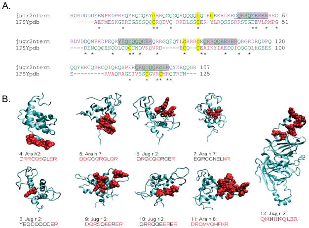
Results: Sequences from the vicilin walnut allergen Jug r 2, which had low PD values to epitopes of the peanut allergen Ara h 2, a 2S albumin, bound to IgE in sera from five patients who reacted to either walnut or peanut or both. A walnut epitope recognized by sera from six patients mapped to a surface-exposed region on a model of the N-terminal pro-region of Jug r 2. This predicted walnut epitope competed for IgE binding to Ara h 2 in serum as well as the known IgE epitope from Ara h 2.
Conclusions: Sequences with low PD value (< 8.5) to known IgE epitopes could contribute to cross-reactivity between allergens. This further validates the PD scoring method for predicting cross-reactive epitopes in allergens.
© 2011 John Wiley & Sons A/S.
Conflict of interest statement
None of the authors have any conflict of interests to disclose regarding this manuscript.
Figures
References
-
- de Leon MP, Drew AC, Glaspole IN, Suphioglu C, O'Hehir RE, Rolland JM. IgE cross-reactivity between the major peanut allergen Ara h 2 and tree nut allergens. Mol Immunol. 2007;44(4):463–71. - PubMed
-
- de Leon MP, Glaspole IN, Drew AC, Rolland JM, O'Hehir RE, Suphioglu C. Immunological analysis of allergenic cross-reactivity between peanut and tree nuts. Clin Exp Allergy. 2003;33(9):1273–80. - PubMed
-
- Teuber S, Beyer K, Comstock S, Wallowitz M. The big eight foods: clinical and epidemiological overview. In: Malecki S, editor. Food Allergy. Washington DC: ASM Press; 2006. pp. 49–79.
-
- Teuber SS, Beyer K. Peanut, tree nut and seed allergies. Current Opinion in Allergy and Clinical Immunology. 2004;4(3):201–203. - PubMed
Publication types
MeSH terms
Substances
Grants and funding
LinkOut - more resources
Full Text Sources
Other Literature Sources
