. 2012 Jan;14(1):140-6.
doi: 10.1111/j.1462-2920.2011.02554.x. Epub 2011 Sep 1.

Microbial rhodopsins on leaf surfaces of terrestrial plants

Affiliations

Microbial rhodopsins on leaf surfaces of terrestrial plants

Nof Atamna-Ismaeel et al. Environ Microbiol. 2012 Jan.

Abstract

The above-ground surfaces of terrestrial plants, the phyllosphere, comprise the main interface between the terrestrial biosphere and solar radiation. It is estimated to host up to 10(26) microbial cells that may intercept part of the photon flux impinging on the leaves. Based on 454-pyrosequencing-generated metagenome data, we report on the existence of diverse microbial rhodopsins in five distinct phyllospheres from tamarisk (Tamarix nilotica), soybean (Glycine max), Arabidopsis (Arabidopsis thaliana), clover (Trifolium repens) and rice (Oryza sativa). Our findings, for the first time describing microbial rhodopsins from non-aquatic habitats, point towards the potential coexistence of microbial rhodopsin-based phototrophy and plant chlorophyll-based photosynthesis, with the different pigments absorbing non-overlapping fractions of the light spectrum.


Figures

Fig. 1
Fig. 1
A phylogenetic tree of rhodopsin amino acid sequences (deduced from the metagenomic data) from the phyllospheres of tamarisk, rice, soybean, Arabidopsis and clover. Following alignment computation (see Experimental procedures), a FastTree version 2.1.1 was used for the calculation of the approximately maximum-likelihood phylogenetic tree using settings for high accuracy. Bootstraps above 60% are shown as black circles at the junctions. PR, proteorhodopsins; HR, halorhodopsins; BR, bacteriorhodopsins; SRI, sensory rhodopsins-I; SRII, sensory rhodopsins-II.
Fig. 2
Fig. 2
Relative abundance of microbial rhodopsins in different metagenomes. MG-RAST (Meyer et al., 2008) accession numbers of the different data sets can be found in Experimental procedures. Abundance was normalized relative to the numbers of rplA, rplC, rplD, rpoA, rpoB and rspJ genes (Frank and Sorensen, 2011) in each environment.
Fig. 3
Fig. 3
Sensory rhodopsins and proton pumps in different environments. Proportions of sensory rhodopsins and rhodopsin proton pumps were calculated only from reads containing the region surrounding the proton acceptor and donor carboxylates at helix C (bacteriorhodopsin positions 85 and 96, respectively); Sargasso Sea (Spudich, 2006) (n = 732), tamarisk (n = 13), soybean (n = 31), rice (n = 8), Arabidopsis (n = 4) and clover (n = 7).
Fig. 4
Fig. 4
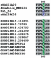
Protein alignment of phyllosphere rhodopsins. Amino acid position 105 is marked with green or blue backgrounds according to the predicted absorption spectra of the rhodopsin pigments. Only the vicinity of amino acid 105 is shown. Examples from confirmed green absorbing proteorhodopsins eBAC31A08 (Béjà et al., 2000), Dokdonia MED134 (Gómez-Consarnau et al., 2007) and confirmed blue absorbing proteorhodopsins PAL-E6 (Béjà et al., 2001), eBAC49C08 (Sabehi et al., 2005) are shown for reference at the top. Names of rhodopsins from the soybean phyllosphere start with SRR and from the tamarisk start with GDOVJJ. Only a subset of the phyllosphere rhodopsins is shown. See Supporting material S1, S2, S3, S4 and S5 for more variations.
Fig. 5
Fig. 5
Absorbance spectra of tamarisk leaves and phyllosphere wash. Absorbance of tamarisk chlorophylls (acetone extract) and of phyllosphere leaf buffer-wash are shown; note the different scales used.

References

    1. Atamna-Ismaeel N, Sabehi G, Sharon I, Witzel K-P, Labrenz M, Jürgens K, et al. Widespread distribution of proteorhodopsins in freshwater and brackish ecosystems. ISME J. 2008;2:656–662. - PubMed
    1. Balashov SP, Imasheva ES, Boichenko VA, Anton J, Wang JM, Lanyi JK. Xanthorhodopsin: a proton pump with a light-harvesting carotenoid antenna. Science. 2005;309:2061–2064. - PMC - PubMed
    1. Ballaré CL, Scopel AL, Sánchez RA. Far-red radiation reflected from adjacent leaves: an early signal of competition in plant canopies. Science. 1990;247:329–332. - PubMed
    1. Beattie GA, Lindow SE. Bacterial colonization of leaves: a spectrum of strategies. Phytopathol. 1999;89:353–359. - PubMed
    1. Béjà O, Aravind L, Koonin EV, Suzuki MT, Hadd A, Nguyen LP, et al. Bacterial rhodopsin: evidence for a new type of phototrophy in the sea. Science. 2000;289:1902–1906. - PubMed

Publication types

Substances