. 2012 Jul;16(7):1533-43.
doi: 10.1111/j.1582-4934.2011.01410.x.

Altered distribution of interstitial cells and innervation in the rat urinary bladder following spinal cord injury

Affiliations

Altered distribution of interstitial cells and innervation in the rat urinary bladder following spinal cord injury

Louise Johnston et al. J Cell Mol Med. 2012 Jul.

Abstract

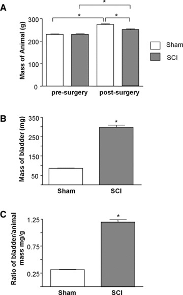
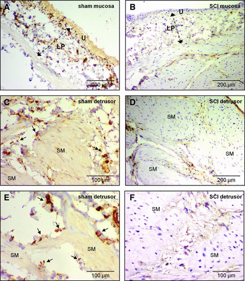
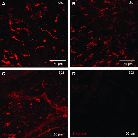
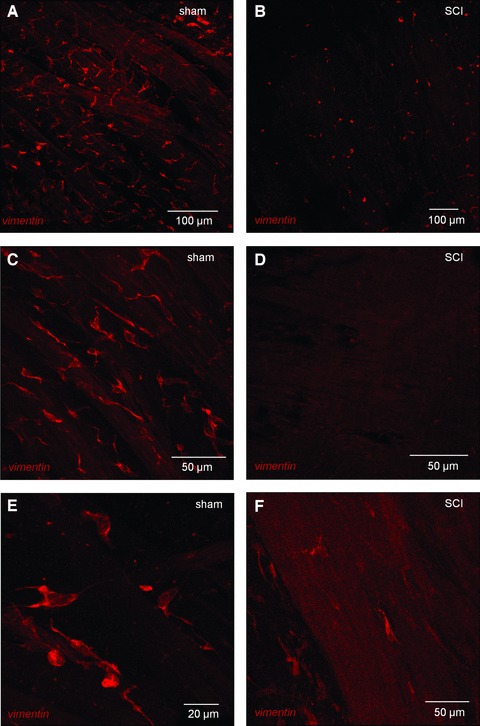
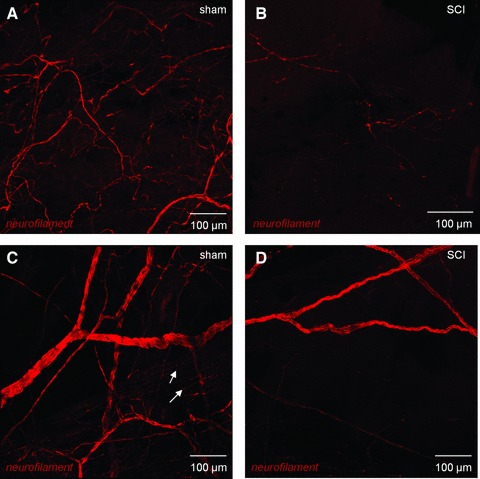
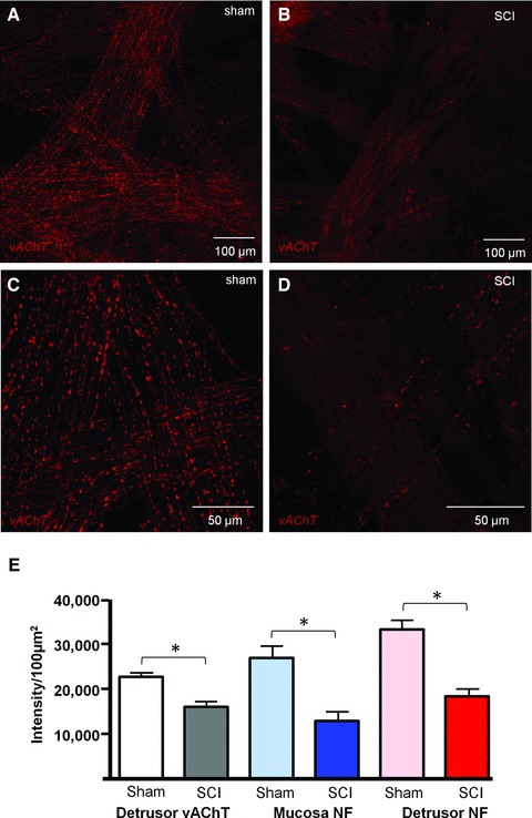
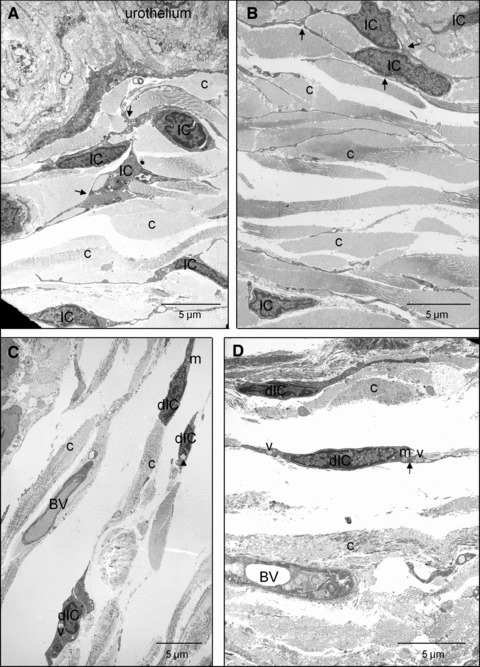
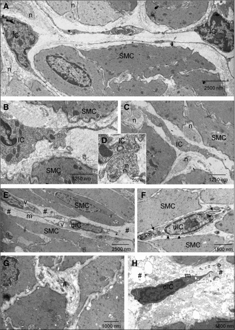
Changes in the distribution of interstitial cells (IC) are reportedly associated with dysfunctional bladder. This study investigated whether spinal cord injury (SCI) resulted in changes to IC subpopulations (vimentin-positive with the ultrastructural profile of IC), smooth muscle and nerves within the bladder wall and correlated cellular remodelling with functional properties. Bladders from SCI (T8/9 transection) and sham-operated rats 5 weeks post-injury were used for ex vivo pressure-volume experiments or processed for morphological analysis with transmission electron microscopy (TEM) and light/confocal microscopy. Pressure-volume relationships revealed low-pressure, hypercompliance in SCI bladders indicative of decompensation. Extensive networks of vimentin-positive IC were typical in sham lamina propria and detrusor but were markedly reduced post-SCI; semi-quantitative analysis showed significant reduction. Nerves labelled with anti-neurofilament and anti-vAChT were notably decreased post-SCI. TEM revealed lamina propria IC and detrusor IC which formed close synaptic-like contacts with vesicle-containing nerve varicosities in shams. Lamina propria and detrusor IC were ultrastructurally damaged post-SCI with retracted/lost cell processes and were adjacent to areas of cellular debris and neuronal degradation. Smooth muscle hypertrophy was common to SCI tissues. In conclusion, IC populations in bladder wall were decreased 5 weeks post-SCI, accompanied with reduced innervation, smooth muscle hypertrophy and increased compliance. These novel findings indicate that bladder wall remodelling post-SCI affects the integrity of interactions between smooth muscle, nerves and IC, with compromised IC populations. Correlation between IC reduction and a hypercompliant phenotype suggests that disruption to bladder IC contribute to pathophysiological processes underpinning the dysfunctional SCI bladder.


Figures

Fig 1
Fig 1
Effect of spinal cord transection on animal and bladder mass. (A) Summary data from sham operated and spinal cord injured animals (N = 21 in each group) showing significant increase in body mass in both groups after the 5-week intervention period and a significant difference in body mass post-surgery between sham and SCI animals. (B) The bladders of SCI animals had significantly greater mass than sham animals post-surgery (N = 21, P < 0.05). (C) Bladder to body mass ratio was significantly higher in the SCI group compared with sham-operated controls (P < 0.05).
Fig 2
Fig 2
Pressure–volume relationships. Pressure–volume curves of ex vivo bladders showing continuous pressure tracings as a function of filling volume. Data are from sham-operated controls (grey) and SCI animals (black). A reduction of slope indicates increased compliance.
Fig 3
Fig 3
Vimentin immunohistochemistry in histological sections. (A) Abundant vimentin-positive cells stained with DAB (brown) in a mucosal section of sham rat bladder. Lamina propria (LP), urothelium (U). Sections were counterstained with haematoxylin. (B) Vimentin-positive cells were less abundant in the lamina propria of SCI bladder. Staining was predominantly in rounded, cell bodies compared with the cellular structures seen in (A). (C, E) Vimentin-positive cells in the detrusor were found on the boundary (arrows) of smooth muscle bundles (SM) and in the inter-bundle spaces of sham bladders. (D, F) In SCI detrusor, vimentin-positive cells were notably less abundant and had a compromised (rounded) cellular morphology. Note the hypertrophied smooth muscle in SCI compared with sham bladders.
Fig 4
Fig 4
Confocal imaging of vimentin-positive cells in lamina propria. (A, B) Whole mount, flat sheet preparations of sham bladder mucosa were labelled with anti-vimentin and imaged with confocal microscopy. All images are reconstructions of optical sections. Networks of vimentin-positive cells were found in the lamina propria. Vimentin-positive cells had bipolar or highly branched stellate morphology. (C) Mucosal tissues from SCI animals also had vimentin-positive cells however, the networks were apparently less well developed than the sham operated controls and cells had a compromised, non-branched morphology. (D) Tissues labelled with only the secondary antibody (primary antibody omitted) had minimal staining.
Fig 5
Fig 5
Vimentin-positive cells in the detrusor of sham and SCI bladders. (A) Whole mount, flat sheet preparations of sham detrusor were labelled with anti-vimentin and imaged with confocal microscopy. All images are reconstructions of optical sections. Vimentin-positive cells formed extensive networks in the detrusor. (C, E) At higher magnification, the immunopositive cells had a highly branched stellate morphology and occupied the spaces between the smooth muscle bundles. Vimentin-positive cells were also found on the boundary of the smooth muscle bundles either bipolar cells, orientated axially or stellate cells spanning two or more bundles as shown in panel C. (B, D, F). Vimentin-positive cells were markedly altered in SCI tissues with only a few patches of cells visible, which generally had lost their stellate morphology.
Fig 6
Fig 6
Confocal imaging of bladder neural networks. (A) Whole mount, flat sheet preparations of mucosa from sham bladders were labelled with anti-neurofilament and imaged with confocal microscopy. All images are reconstructions of optical sections. A neural plexus was observed in these sections comprising larger nerves, which bifurcated to increasingly smaller fibres. (B) In SCI animals, the neural plexus was significantly disrupted with an apparent reduction in nerves. (C) The detrusor region of sham control bladders was densely innervated with large nerve trunks subdividing into increasingly smaller nerves, which became varicosities (arrows) within the smooth muscle bundles. Mucosal tissues from SCI animals also had strikingly compromised neuronal distribution with only occasional patches of immunopositive nerves. (D) In SCI detrusor, the innervation was comparatively sparse with only patchy areas of nerves visible.
Fig 7
Fig 7
Confocal imaging of cholinergic nerves. (A, C) Whole mount, flat sheet preparations of sham detrusor were labelled with anti-vesicular acetylcholinetransferase (vAChT) and imaged with confocal microscopy. All images are reconstructions of optical sections. A dense cholinergic innervation was clear within the detrusor. (B, D) SCI detrusor had marked reduction in vAChT varicosities. (E) Summary graph of the effect of spinal cord injury on vAChT-positive nerves in the detrusor (n = 16 micrographs from N = 2 bladders, Sham and SCI groups), neurofilament-positive nerves in the mucosa (n = 18, N = 3 sham; n = 14, N = 3 SCI) and detrusor (n = 24, N = 3 sham; n = 25, N = 3 SCI). The asterisk indicates P < 0.05.
Fig 8
Fig 8
Transmission electron micrographs of lamina propria IC in sham and SCI bladders. (A, B) The lamina propria region of sham bladders had abundant cells with the ultrastructural properties of IC. Cell–cell interactions are shown by arrows and collagen was abundant (c). The base of the urothelium is visible. (C, D) After SCI, damaged IC (dIC) were apparent, although their branched process and cell–cell contacts were less extensive than sham controls. dIC typically had cytoplasmic vacuolisation (v), swollen endoplasmic reticulum (arrows), apparent collapse of the cytoskeleton in the perinuclear area (arrowheads) and some swollen mitochondria (m). Blood vessels are visible (BV).
Fig 9
Fig 9
Transmission electron micrographs of detrusor IC in sham and SCI bladders (A–C) Cells with ultrastructural characteristics of IC were present in sham detrusors. These had cytoplasmic branches and were close to smooth muscle cells (SMC). Nerve (n) varicosities were abundant and were in close contact with both SMC and IC. (D) Magnification of the boxed area in B showing synapse-like contact between an IC and a nerve ending. (E–H) After SCI, damaged IC (dIC) were present with evidence of damage to branched processes and cell–cell contacts. dIC typically had cytoplasmic vacuolisation (v), swollen endoplasmic reticulum (arrows), apparent collapse of the cytoskeleton in the perinuclear area (arrowheads) and some swollen mitochondria (m). Areas of cellular debris were abundant (#). SMC exhibited a lesser degree of degradation.

Similar articles

Cited by

References

    1. Abrams PH, Dunn M, George N. Urodynamic findings in chronic retention of urine and their relevance to results of surgery. Br Med J. 1978;2:1258–60. - PMC - PubMed
    1. Kalejaiye O, Speakman MJ. Management of acute and chronic retention in men. Eur Urol. 2009;8:523–9.
    1. Cruz C, Cruz F. Spinal cord injury and bladder dysfunction: new ideas about an old problem. Scientific World J. 2011;11:214–34. - PMC - PubMed
    1. McCloskey KD. Interstitial cells of Cajal in the urinary tract. Handb Exp Pharmacol. 2011a;202:233–54. - PubMed
    1. Hashitani H, Lang RJ. Functions of ICC-like cells in the urinary tract and male genital organs. J Cell Mol Med. 2010;14:1199–211. - PMC - PubMed

Publication types

MeSH terms