. 2011 Nov;15(11):2539-51.
doi: 10.1111/j.1582-4934.2011.01417.x.

Cardiomyocytes derived from human embryonic and induced pluripotent stem cells: comparative ultrastructure

Affiliations

Cardiomyocytes derived from human embryonic and induced pluripotent stem cells: comparative ultrastructure

Mihaela Gherghiceanu et al. J Cell Mol Med. 2011 Nov.

Abstract

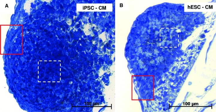
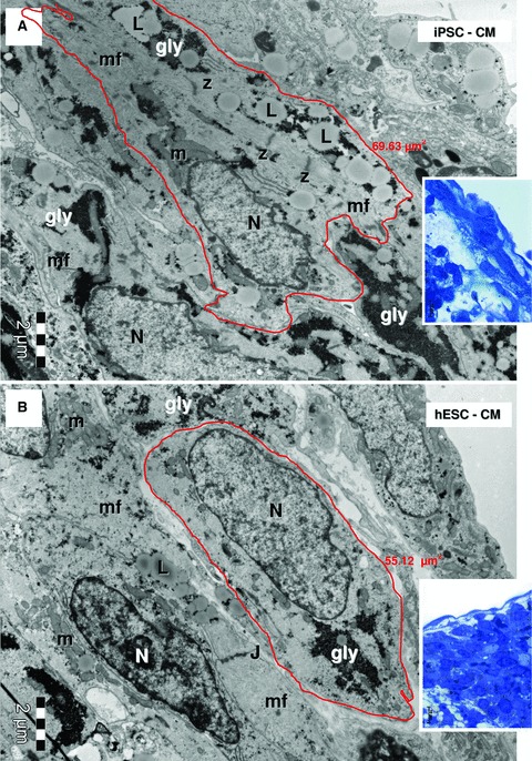
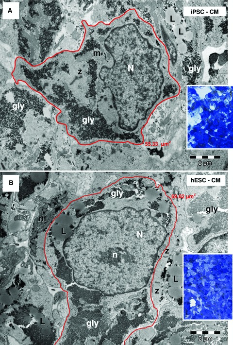
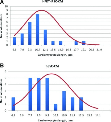
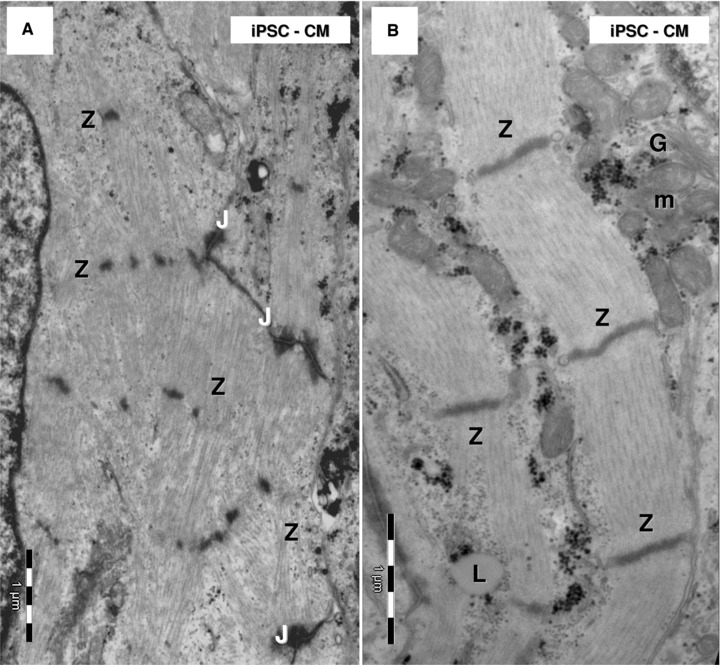
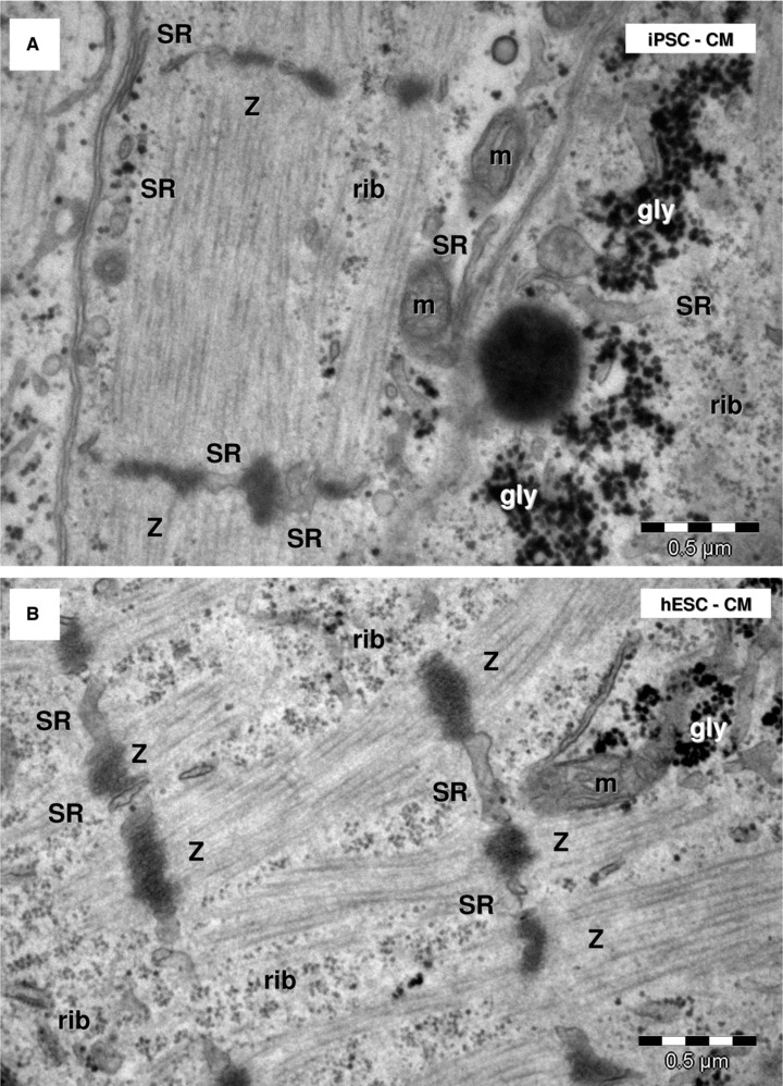
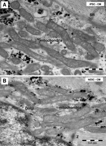
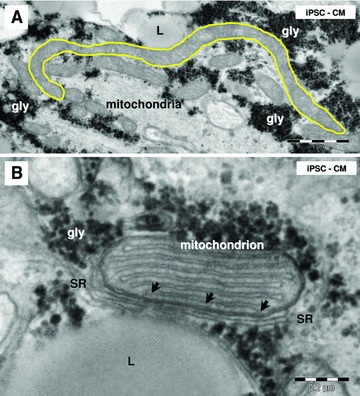
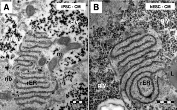
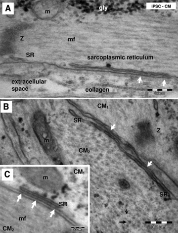
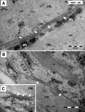
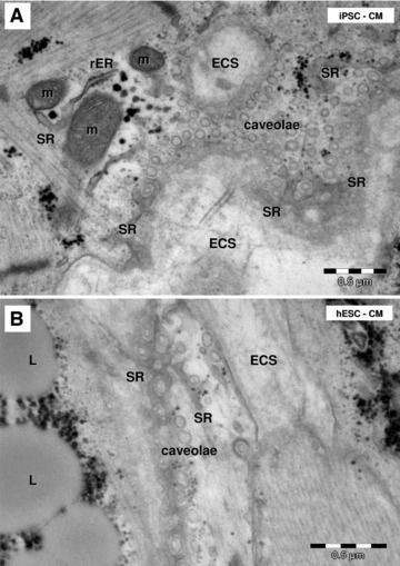
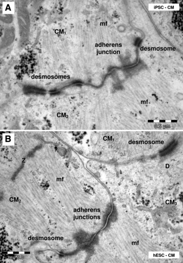
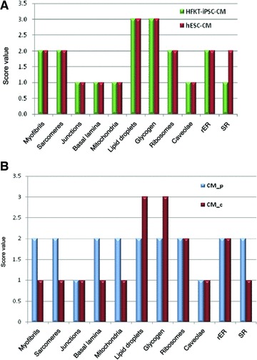
Induced pluripotent stem cells (iPSC) are generated from fully differentiated somatic cells that were reprogrammed into a pluripotent state. Human iPSC which can be obtained from various types of somatic cells such as fibroblasts or keratinocytes can differentiate into cardiomyocytes (iPSC-CM), which exhibit cardiac-like transmembrane action potentials, intracellular Ca(2+) transients and contractions. While major features of the excitation-contraction coupling of iPSC-CM have been well-described, very little is known on the ultrastructure of these cardiomyocytes. The ultrastructural features of 31-day-old (post-plating) iPSC-CM generated from human hair follicle keratinocytes (HFKT-iPSC-CM) were analysed by electron microscopy, and compared with those of human embryonic stem-cell-derived cardiomyocytes (hESC-CM). The comparison showed that cardiomyocytes from the two sources share similar proprieties. Specifically, HFKT-iPSC-CM and hESC-CM, displayed ultrastructural features of early and immature phenotype: myofibrils with sarcomeric pattern, large glycogen deposits, lipid droplets, long and slender mitochondria, free ribosomes, rough endoplasmic reticulum, sarcoplasmic reticulum and caveolae. Noteworthy, the SR is less developed in HFKT-iPSC-CM. We also found in both cell types: (1) 'Ca(2+)-release units', which connect the peripheral sarcoplasmic reticulum with plasmalemma; and (2) intercellular junctions, which mimic intercalated disks (desmosomes and fascia adherens). In conclusion, iPSC and hESC differentiate into cardiomyocytes of comparable ultrastructure, thus supporting the notion that iPSC offer a viable option for an autologous cell source for cardiac regenerative therapy.


Figures

Fig 1
Fig 1
Electrophysiological features of cardiomyocytes derived from iPSC generated from human plucked hair follicle keratinocytes (HFKT-iPSC-CM; clone KTN3). (A) Extracellular electrograms recorded by means of MEA data acquisition system from a spontaneously contracting embryoid body (EB). (B) Representative action potential recordings from a spontaneously contracting EB (clone KTN3), demonstrating the pacemaker activity of the HFKT-iPSC-CM.
Fig 2
Fig 2
Light microscopy of semi-thin sections. Toluidine blue staining of resin-embedded embryoid bodies of HFKT-iPSC (A) and hESC (B). The red squared marked areas in the periphery of EBs in (A) and (B) have the corresponding light and electron microscopy in Figure 3. Light and electron microscopy of areas marked with white dashed lines in (A) and (B) are detailed in Figure 4.
Fig 3
Fig 3
Electron micrographs of the EBs periphery (red marked areas in Fig. 1A and B, higher magnification of blue sections in the corresponding insets) show ellipsoid cardiomyocytes derived from HFK-iPSC (A) and hESC (B). Myofilaments (mf) organized in distinct sarcomeric structures delineated by Z-bands. Lipid droplets (L), glycogen masses (gly) and clusters of mitochondria (m) are visible in cytoplasm of cardiomyocytes. Small junctions (J) connect the cardiomyocytes. N: nucleus. The measured areas of red outlined cardiomyocytes are 69.63 μm2 for HFK-iPSC-CM (A) and 55.12 μm2 for hESC-CM (B).
Fig 4
Fig 4
Electron micrographs of the EBs centre (white marked areas in Fig. 2A and B) show cardiomyocytes with irregular contour derived from HFK-iPSC (A) and hESC (B). Insets show corresponding blue sections. Cardiomyocytes have myofibrils (mf), Z-bands, lipid droplets (L), glycogen (gly), mitochondria (m). N: nucleus; n: nucleolus. The measured areas of red outlined cardiomyocytes are 55.33 μm2 for HFK-iPSC-CM (A) and 69.62 μm2 for hESC-CM (B).
Fig 5
Fig 5
Histograms for measured lengths of HFK-iPSC-CM (A) and hESC-CM (B). The red line represents the ideal situation. In each group, 28 cardiomyocytes were analysed. The analysis included three EBs from each group.
Fig 6
Fig 6
Electron microscopy of HFK-iPSC-CM. Note highly organized myofilaments which form myofibrils (A) or myofibrils lacking alignment (B) in HFK-iPSC-CM. (A) Z-like dense structures connect bundles of myofilaments. (B) Z-bands clearly show sarcomere limits along myofibrils. J: junctions; L: lipid droplet; m: mitochondria; G: Golgi system.
Fig 7
Fig 7
Electron microscopy shows myofibrils with sarcomeric pattern in HFK-iPSC-CM (A) and hESC-CM (B). Sarcoplasmic reticulum cisternae (SR) are visible between Z-bands. m: mitochondria; rib: free ribosomes; gly: glycogen granules.
Fig 8
Fig 8
Electron microscopy shows clusters of long and slender mitochondria in HFK-iPSC-CM (A) and hESC-CM (B). SR: sarcoplasmic reticulum; gly: glycogen granules; Z: Z band.
Fig 9
Fig 9
Electron microscopy of iPSC-CM mitochondria. (A) Note a long mitochondrion (yellow outlined; 6 μm). (B) Small dense structures (arrows) connect sarcoplasmic reticulum (SR) with a mitochondrion. L: lipid droplets; gly: glycogen.
Fig 10
Fig 10
Electron microscopy shows conspicuous rough endoplasmic reticulum (rER) in HFK-iPSC-CM (A) and hESC-CM (B). m: mitochondria; rib: ribosomes; L: lipid droplet; gly: glycogen.
Fig 11
Fig 11
Electron micrographs (A–C) show peripheral couplings or ‘Ca2+ release units’ in HFK-iPSC-CM visible as small dense structures (white arrows) between peripheral sarcoplasmic reticulum (SR) and plasmalemma. Longitudinally (A) and transversally (B) sectioned myofilaments. Thin (small black arrow) and thick (thick black arrow) myofilaments are visible in the transverse section (B). Z: Z bands; gly: glycogen; mf: myofilaments; m: mitochondria; CM1 and CM2: indicates that these are different adjacent cardiomyocytes.
Fig 12
Fig 12
Electron micrographs (A–C) show ‘Ca2+ release units’ (white arrows) connecting peripheral sarcoplasmic reticulum (SR) and plasmalemma in hESC-CM. gly: glycogen; mf: myofibrils; m: mitochondria; CM1 and CM2: indicates that these are different adjacent cardiomyocytes.
Fig 13
Fig 13
Electron microscopy shows caveolae on plasmalemma of HFK-iPSC-CM (A) and hESC-CM (B). Tangent sections of cardiomyocytes plasmalemma show the caveolae surrounded by the peripheral sarcoplasmic reticulum (SR). m: mitochondria; rER: rough endoplasmic reticulum; L: lipid droplets; ECS: extracellular space.
Fig 14
Fig 14
Electron microscopy shows desmosomes and adheres junctions connecting HFK-iPSC-CM (A) and hESC-CM (B). CM1–CM3: different cardiomyocytes; Z: Z-bands; mf: myofilaments.
Fig 15
Fig 15
(A) Comparative representation of semi-quantitative assessment of the ultrastructural features of HFK-iPSC-CM and hESC-CM. (B) Comparative representation of semi-quantitative assessment of the ultrastructural features of peripheral (CM_p) and central (CM_c) located cardiomyocytes into the embryoid bodies. rER: rough endoplasmic reticulum; SR: sarcoplasmic reticulum.

References

    1. Takahashi K, Yamanaka S. Induction of pluripotent stem cells from mouse embryonic and adult fibroblast cultures by defined factors. Cell. 2006;126:663–76. - PubMed
    1. Takahashi K, Tanabe K, Ohnuki M, et al. Induction of pluripotent stem cells from adult human fibroblasts by defined factors. Cell. 2007;131:861–72. - PubMed
    1. Germanguz I, Sedan O, Zeevi-Levin N, et al. Molecular characterization and functional properties of cardiomyocytes derived from human inducible pluripotent stem cells. J Cell Mol Med. 2011;15:38–51. - PMC - PubMed
    1. Novak A, Shtrichman R, Germanguz I, et al. Enhanced reprogramming and cardiac differentiation of human keratinocytes derived from plucked hair follicles, using a single excisable lentivirus. Cell Reprogramm. 2010;12:665–78. - PubMed
    1. Fujiwara M, Yan P, Otsuji TG, et al. Induction and enhancement of cardiac cell differentiation from mouse and human induced pluripotent stem cells with cyclosporine-A. PLoS One. 2011;6:e16734. 10.1371/journal.pone.0016734. - PMC - PubMed

Publication types

MeSH terms