Review
. 2011 Sep;243(1):74-90.
doi: 10.1111/j.1600-065X.2011.01049.x.

dsRNA sensors and plasmacytoid dendritic cells in host defense and autoimmunity

Affiliations
Review

dsRNA sensors and plasmacytoid dendritic cells in host defense and autoimmunity

Yaming Wang et al. Immunol Rev. 2011 Sep.

Abstract

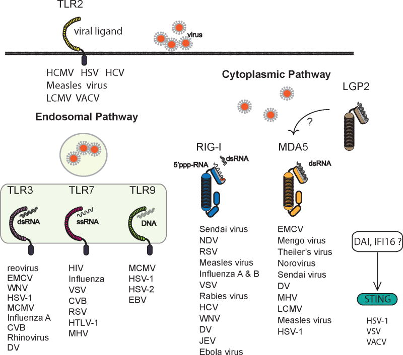
The innate immune system detects viruses through molecular sensors that trigger the production of type I interferons (IFN-I) and inflammatory cytokines. As viruses vary tremendously in size, structure, genomic composition, and tissue tropism, multiple sensors are required to detect their presence in various cell types and tissues. In this review, we summarize current knowledge of the diversity, specificity, and signaling pathways downstream of viral sensors and ask whether two distinct sensors that recognize the same viral component are complementary, compensatory, or simply redundant. We also discuss why viral sensors are differentially distributed in distinct cell types and whether a particular cell type dominates the IFN-I response during viral infection. Finally, we review evidence suggesting that inappropriate signaling through viral sensors may induce autoimmunity. The picture emerging from these studies is that disparate viral sensors in different cell types form a dynamic and integrated molecular network that can be exploited for improving vaccination and therapeutic strategies for infectious and autoimmune diseases.


Conflict of interest statement

The authors have no conflicts of interest to declare.

Figures

Fig. 1
Fig. 1. Diversity and specificity of viral sensors
Multiple viral sensors detect invading viruses. Genetic loss-of-function experiments have revealed the specificity of viral sensors in numerous viral infections. TLR2 is expressed on the cell surface and detects Human cytomegalovirus (HCMV), HSV-1, Hepatitis C virus (HCV), Measles virus, LCMV, and VACV virus (–20). TLR3, TLR7, and TLR9 are located in endosomes. TLR3 recognizes RNA and DNA viruses such as reovirus (25), EMCV (123), West Nile virus (WNV) (234, 235), HSV-1 (35), MCMV (236), Influenza A (40), CVB (128), rhinovirus (237, 238), and Dengue virus (DV) (239). TLR7 recognizes ssRNA viruses such as human immunodeficiency virus (HIV) (240), Influenza virus (41, 42), VSV (43), CVB (241) and respiratory syncytial virus (RSV) (242), human T-cell leukemia virus (HTLV-1) (257) and mouse hepatitis virus (MHV) (258). TLR9 recognizes DNA viruses such as MCMV (243), HSV-1 (244), HSV-2 (245) and Epstein-Barr virus (EBV) (246). RIG-I and MDA5 sense replicating viruses in the cytosol. RIG-I recognizes a series of ssRNA viruses including paramyxoviruses such as Sendai virus (71, 247), Newcastle disease virus (NDV) (71, 247), RSV (248) and Measles virus (249, 250); Orthomyxovirus (Influenza A and B viruses (71)); Rhabdovirus (VSV (71, 247), Rabies virus (251)); Flavivirus (HCV (74), WNV (252), DV (239), Japanese encephalitis virus (JEV) (71)); and Filovirus (Ebola virus (95)). MDA5 detects picornaviruses such as EMCV, Mengo virus, and Theiler virus (71, 72), as well as calicivurses, such as norovirus (253). MDA5 has also been shown to recognize Sendai virus (75), DV (239), mouse hepatitis virus (MHV) (254), LCMV (233), Measles virus (249) and HSV-1 (255). DNA sensors such as DAI and IFI16 recognize cytosolic DNA viruses in a STING-dependent manner (99, 100, 103-106). STING is also involved in the recognition of RNA viruses such as VSV (105, 106).
Fig. 2
Fig. 2. Hematopoietic and stromal contributions to poly(I:C)-mediated immunity
(A). Poly(I:C) promotes CD4+ and CD8+ T cell immunity through the production of IFN-I and other cytokines. Poly(I:C)-mediated IFN-I responses are dependent on stromal expression of MDA5. However, adjuvant effects of poly(I:C) on CD4+ T cell immunity require MDA5 expression in both hematopoietic and stromal compartments. Poly(I:C) triggers the TLR3 pathway in CD8α+ DCs to promote cross-presentation to CD8+ T cells while signaling through MDA5 pathway in stromal cells promotes CD8+ T cell survival and memory formation. (B). Poly(I:C) promotes NK cell activation through the production of IFN-I and IL-12. NK cell activation via IFN-I depends on MDA5-expressing radio-resistant stromal cells while TLR3+ hematopoietic cells promote NK cell activation through IL-12 secretion. The MDA5 pathway plays a dominant role in NK cell responses to poly(I:C) while the TLR3 pathway has a secondary effect.
Fig. 3
Fig. 3. Cellular sources of IFN-I during viral infections
TLR7- and TLR9-expressing pDCs detect RNA and DNA viruses and provide an initial source of IFN-I. However, pDC-mediated IFN-I production is limited and transient, indicating that the majority IFN-I required for controlling viral replication is derived from additional cellular sources such as cDCs, macrophages, monocytes, B cells, and stromal cells that utilize TLRs, RLRs, and DNA sensors for viral detection. The expression of RLRs and DNA sensors is often low but highly inducible in response of IFN-I, suggesting that initial IFN-I by pDCs may be essential for RLR- and DNA sensor-mediated responses.
Fig. 4
Fig. 4. MDA5 and IFN-I in virus-induced and autoimmune diabetes
Viruses such as EMCV and CVB cause diabetes in mice. Polymorphisms in MDA5, a sensor for β-cell tropic viruses such as CVB and EMCV-D, have been linked to the development of human T1D. In virus-induced diabetes, activation of the MDA5 pathway in DCs and macrophages triggers IFN-I responses, which inhibits viral replication and prevents βcell injury. On the other hand, in autoimmune diabetes, MDA5 signaling might elicit excessive IFN-I production that could induce apoptosis of β-cells and promote presentation of β-cell antigens to autoreactive T cells by islet-resident DCs (256). IFN-I is also capable of activating autoreactive T cells directly in an antigen non-specific fashion.

References

    1. Pestka S, Krause CD, Walter MR. Interferons, interferon-like cytokines, and their receptors. Immunol Rev. 2004;202:8–32. - PubMed
    1. Malathi K, et al. A transcriptional signaling pathway in the IFN system mediated by 2′-5′-oligoadenylate activation of RNase L. Proc Natl Acad Sci USA. 2005;102:14533–14538. - PMC - PubMed
    1. Malathi K, Dong B, Gale M, Jr, Silverman RH. Small self-RNA generated by RNase L amplifies antiviral innate immunity. Nature. 2007;448:816–819. - PMC - PubMed
    1. Barber GN, Wambach M, Wong ML, Dever TE, Hinnebusch AG, Katze MG. Translational regulation by the interferon-induced double-stranded-RNA-activated 68-kDa protein kinase. Proc Natl Acad Sci USA. 1993;90:4621–4625. - PMC - PubMed
    1. Theofilopoulos AN, Baccala R, Beutler B, Kono DH. Type I interferons (alpha/beta) in immunity and autoimmunity. Annu Rev Immunol. 2005;23:307–336. - PubMed

Publication types