Review
. 2011 Sep;243(1):191-205.
doi: 10.1111/j.1600-065X.2011.01040.x.

Innate and adaptive immune responses to cell death

Affiliations
Review

Innate and adaptive immune responses to cell death

Kenneth L Rock et al. Immunol Rev. 2011 Sep.

Abstract

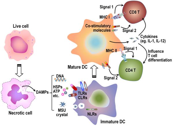
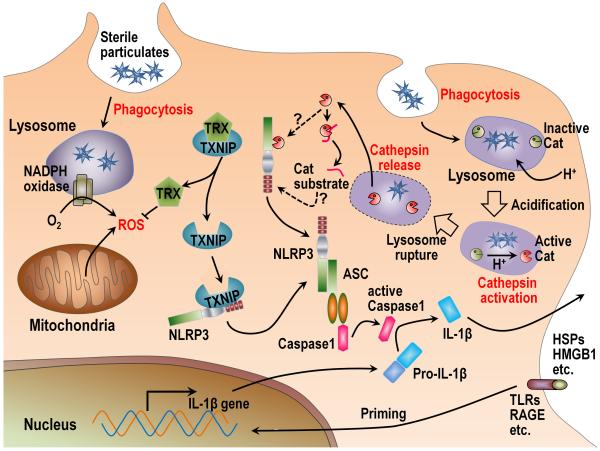
The immune system plays an essential role in protecting the host against infections and to accomplish this task has evolved mechanisms to recognize microbes and destroy them. In addition, it monitors the health of cells and responds to ones that have been injured and killed, even if this occurs under sterile conditions. This process is initiated when dying cells expose intracellular molecules that can be recognized by cells of the innate immune system. As a consequence of this recognition, dendritic cells are activated in ways that help to promote T-cell responses to antigens associated with the dying cells. In addition, macrophages are stimulated to produce the cytokine interleukin-1 that then acts on radioresistant parenchymal cells in the host in ways that drive a robust inflammatory response. In addition to dead cells, a number of other sterile particles and altered physiological states can similarly stimulate an inflammatory response and do so through common pathways involving the inflammasome and interleukin-1. These pathways underlie the pathogenesis of a number of diseases.


Figures

Fig. 1
Fig. 1. Cells contain an endogenous adjuvant activity that augments the priming of CD8+ T-cell responses
A limiting amount of antigen (5 μg ovalbumin-conjugated beads) was co-injected into mice together with either 105, 104, or103 mitomycin C-treated syngeneic GL261 cells. Seven days later, splenocytes were harvested and stimulated with ovalbumin-transfected EG7 cells. CTL activity was measured against EG7 cells 5 days later in a 51Cr release assay. E:T ratio= 33:1. The data show that the GL261 cells, which lack antigen, contain an activity that markedly augments the priming of CD8+ T-cell responses to the co-injected antigen.
Fig. 2
Fig. 2. Adjuvant effect of DAMPs in stimulating dendritic cells
When cells undergo necrosis intracellular DAMPs (such as DNA, HSPs, MSU, etc.) are released into the extracellular milieu. Some of these DAMPs can act as adjuvants to stimulate dendritic cells (DCs), through pattern recognition receptors, such as TLRs, CLRs, or NLRs, or other receptors to increase the expression of MHC molecules, co-stimulatory ligands, and cytokines. These mature DC can then optimally activate T cells and direct their differentiation. HSPs, heat-shock proteins; MSU, monosodium urate crystal; TLRs, Toll-like receptors; CLRs, C-type lectin receptors; NLRs, NOD-like receptors.
Fig. 3
Fig. 3. Role of IL-1 and TNFα in cell death-induced inflammatory responses
Wildtype (WT) C57BL/6 and (A) IL-1αβ double-deficient or (B) TNFα-deficient mice were injected intraperitoneally with 30 million heat-shocked necrotic EL4 cells. Total neutrophil number in the peritoneal cavity was determined 14 h after stimulation. The data are combined results of three experiments with values from individual mice displayed along with means ± SEM. *p<0.05, ***p<0.0001 versus WT group. The data shown that TNFα can make some contribution to the cell death-induced inflammatory responses; notably this contribution is less than from IL-1.
Fig. 4
Fig. 4. Analysis of the cells required for the generation of cell death-induced inflammation
Wildtype (WT, FVB/N) or CD11b-DTR-Tg mice were injected intravenously with 500 ng of diphtheria toxin (500 ng); this procedure depletes macrophages in the transgenic mice. The treated animals were then reconstituted with F4/80+ macrophages (MPs) from thioglycollate-elicited peritoneal cells or CD11c+ dendritic cells (DCs) from spleens of Flt3L-treated mice (C57BL/6 mice injected with Flt3L-transduced cells). Neutrophil number in the peritoneum was determined 14 h after intraperitoneal injection of heat-shocked necrotic EL4 cells or PBS and displayed as the mean ± SEM. The data show that loss of CD11b+ cells in the host markedly inhibits inflammation to dead cells and that these responses can be reconstituted by the adoptive transfer of MPs or DCs.
Fig. 5
Fig. 5. Mechanisms of NLRP3 inflammasome activation by sterile stimuli
There are two distinct mechanisms proposed as to how NLRP3 inflammasomes are activated by sterile particles. The first model suggests that after phagocytosis, the particles stimulate ROS from NADPH oxidase and/or mitochondria. The increased level of ROS in the cytosol is then sensed by thioredoxin (TRX) and causes its dissociation from thioredoxin-binding protein (TXNIP). The released TXNIP then binds and activates NLRP3 through interaction with LRR and NATCH domains of NLRP3. The second model also requires phagocytosis of the sterile particles. In this pathway phagosomes acidify and the drop in pH causes Cathepsin (Cat) activation. Through some unknown mechanism, some of the particulate-containing phagosomes rupture and release their contents into the cytosol. This vacuolar rupture is somehow sensed by NLRP3, possibly by binding a cleavage product of activated cathepsins or by the cathepsins cleaving NLRP3 in a way that activates it. NLRP3 associates with ASC and pro-Caspase 1 to form the inflammasome and cleaves the Caspase 1 zymogen into its active form. Active Caspase 1 then cleaves pro-IL-1β into active IL-1β. In most cases, the synthesis of pro-IL-1β is induced by “priming”, which requires the stimulation of pattern recognition receptors or cytokines to stimulate transcription of pro-IL-1β.

References

    1. Rock KL, Kono H. The inflammatory response to cell death. Annu Rev Pathol. 2008;3:99–126. - PMC - PubMed
    1. Kono H, Rock KL. How dying cells alert the immune system to danger. Nat Rev Immunol. 2008;8:279–289. - PMC - PubMed
    1. Matzinger P. Tolerance, danger, and the extended family. Annu Rev Immunol. 1994;12:991–1045. - PubMed
    1. Matzinger P. The danger model: a renewed sense of self. Science. 2002;296:301–305. - PubMed
    1. Majno G, Joris I. Cells, tissues, and disease: principles of general pathology. 2nd ed. Oxford University Press; New York; Oxford: 2004.

Publication types