. 2011 Sep;61(9):528-35.
doi: 10.1111/j.1440-1827.2011.02695.x. Epub 2011 Aug 1.

Macrophage polarization in the maculae of age-related macular degeneration: a pilot study

Affiliations

Macrophage polarization in the maculae of age-related macular degeneration: a pilot study

Xiaoguang Cao et al. Pathol Int. 2011 Sep.

Abstract

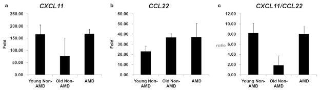
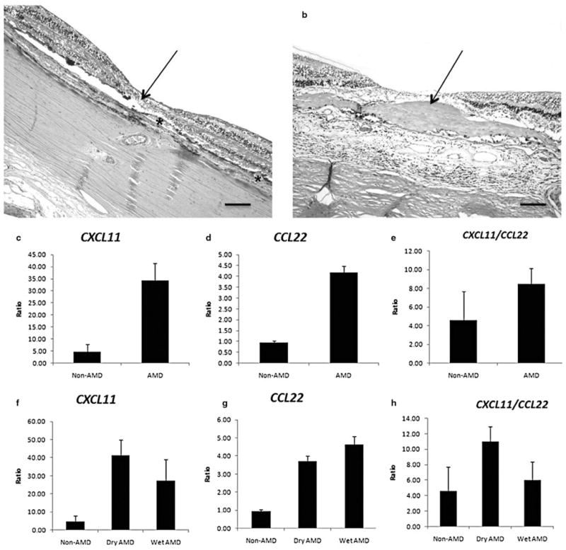
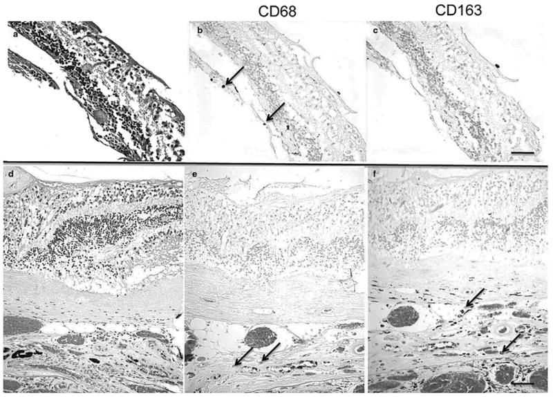
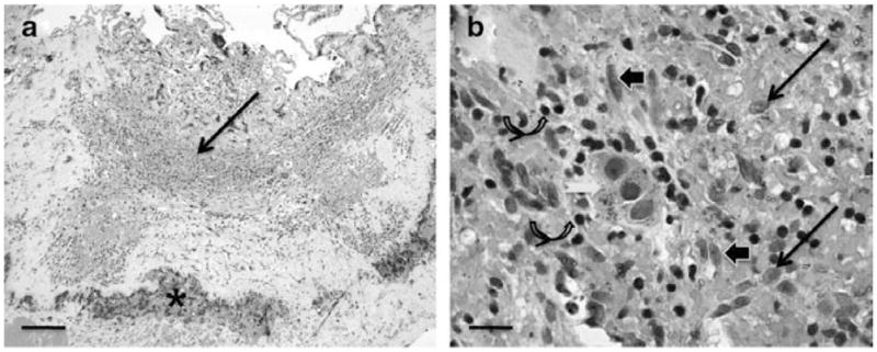
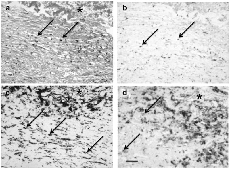
Macrophages can be polarized to exhibit either pro-inflammatory M1 or pro-angiogenic M2 phenotypes, but have high phenotypic plasticity. This pilot study investigated macrophage polarization in the macular retina and choroid of age-related macular degeneration (AMD) and non-AMD subjects, as well as in AMD choroidal neovascular membranes (CNVM). All specimens were evaluated for routine histopathology. Quantitative real-time polymerase chain reaction for representative M1 (CXCL11) and M2 (CCL22) transcripts were performed on macular choroidal trephines (MCT) of 19 AMD and nine non-AMD eye bank eyes, on the microdissected macular retinal cells from the archived slides of five geographic atrophic AMD, five exudative/neovascular AMD, and eight normal autopsied eyes, and on microdissected inflammatory cells from two surgically removed CNVM that did not respond to anti-vascular endothelial growth factor (VEGF) therapy. High M2-chemokine transcript and a low ratio of M1 to M2 chemokine transcript were found in aging non-AMD MCT. Advanced AMD maculae had a higher M1 to M2 chemokine transcript ratio compared to normal autopsied eyes. Macrophages in the two CNVM of patients unresponsive to anti-VEGF therapy were polarized toward either M1 or M2 phenotypes. The number of M2 macrophages was increased compared to M1 macrophages in normal aging eyes. A pathological shift of macrophage polarization may play a potential role in AMD pathogenesis.


Figures

Figure 1
Figure 1
Gene expression of macrophage chemokines for (a) CXCL11 (b) CCL22 and (c) ratio of CXCL11/CCL22 in the macular choroidal trephinates. Young non-age-related macular degeneration (AMD) subgroup (n = 7): <70-year-old non-AMD subjects; old non-AMD subgroup (n = 2): >70-year-old non-AMD subjects; AMD subgroup (n = 19): AMD patients. All values are presented as mean ± SE.
Figure 2
Figure 2
Histology for (a) Geographic atrophy age-related macular degeneration (aAMD) and (b) neovascular/exudative AMD (eAMD), and the relative gene expression ratios between the macular lesions and peripheral retinas of macrophage chemokines for CXCL11 (c and f), CCL22 (d and g) and ratio of CXCL11/CCL22 (e and h) in the archived slides. (a) There is loss of photoreceptor cells (arrow) including the outer and inner segments of the photoreceptors and the outer nuclear layer, abnormal retinal pigment epithelium with hypertrophy and hypotrophy, and many drusen (asterisks) at the fovea and perifovea. (b) Photoreceptor cells and normal RPE are replaced with a thick layer of fibrovascular tissue (arrow) in the maculae. (ch) Measurable relative expression ratios of chemokines are plotted and compared in bar grafts. Non-AMD group (n = 2): age-matched non-AMD subjects; AMD group (n = 10): AMD patients; Dry AMD (n = 5): aAMD; Wet AMD (n = 5): (eAMD). All values are presented as mean ± SE (a and b, HE, original magnifications: ×100, scale bar, 80 μm).
Figure 3
Figure 3
Histopathology of the macular lesions from two age-related macular degeneration patients (AMD; autopsied eyes). (a) A geographic atrophy AMD maculae shows loss of photoreceptors. (b) There is CD68+ macrophage infiltration (arrows). (c) CD163+ cell is not found. (d) A neovascular/exudative AMD maculae shows a thick layer of choroidal neovascularization (CNV). (e) Few CD68+ macrophages (arrows) are indicated in the lesion. (f) More CD163+ macrophages (arrows) are observed in the CNV lesion. (a and d, HE; b, c, e, f, avidin-biotin-complex immunoperoxidase; original magnifications, ac, ×200, scale bar, 60 μm; df, ×100, scale bar, 80 μm).
Figure 4
Figure 4
Histopathology of the choroidal neovascular membrane (CNVM) specimens from the first age-related macular degeneration patient who was unresponsive to bevacizumab therapy. (a) The CNVM consists of moderate monocytic infiltrate (arrow) and small hemorrhages (asterisk); (b) Higher magnification showing many macrophages (arrows), lymphocytes (curved arrows), fibroblasts (arrowheads) and degenerated RPE cells (white arrow). (HE; original magnifications: a, ×50, scale bar, 100 μm; b, ×400, scale bar, 50 μm).
Figure 5
Figure 5
Histopathology of the choroidal neovascular membrane (CNVM) specimens from the second age-related macular degeneration patient who was unresponsive to ranibizumab therapy. (a) The CNVM consists of mild monocytic infiltration (arrows) and hemorrhages (asterisk); (b) Cells in the CNVM express VEGF (arrows); (c and d) Infiltrating cells are CD68+ (c) and CD163+ (d) macrophages. (Original magnifications: ×100, scale bar, 60 μm; a, hematoxylin, bd, avidin biotin-complex immunoperoxidase).

References

    1. World Health Organization. Visual impairment and blindness. Geneva, Switzerland: WHO; 2009. [Accessed 9 October 2010]. Available from: http://www.who.int/mediacentre/factsheets/fs282/en/index.html.
    1. Patel M, Chan CC. Immunopathological aspects of age-related macular degeneration. Semin Immunopathol. 2008;30:97–110. - PMC - PubMed
    1. Dastgheib K, Green WR. Granulomatous reaction to Bruch’s membrane in age-related macular degeneration. Arch Ophthalmol. 1994;112:813–8. - PubMed
    1. Ding X, Patel M, Chan CC. Molecular pathology of age-related macular degeneration. Prog Retin Eye Res. 2009;28:1–18. - PMC - PubMed
    1. Penfold PL, Killingsworth MC, Sarks SH. Senile macular degeneration: The involvement of immunocompetent cells. Graefes Arch Clin Exp Ophthalmol. 1985;223:69–76. - PubMed

Publication types

MeSH terms