. 2011 Aug 31:12:48.
doi: 10.1186/1471-2091-12-48.

Application of Celluspots peptide arrays for the analysis of the binding specificity of epigenetic reading domains to modified histone tails

Affiliations

Application of Celluspots peptide arrays for the analysis of the binding specificity of epigenetic reading domains to modified histone tails

Ina Bock et al. BMC Biochem. .

Abstract

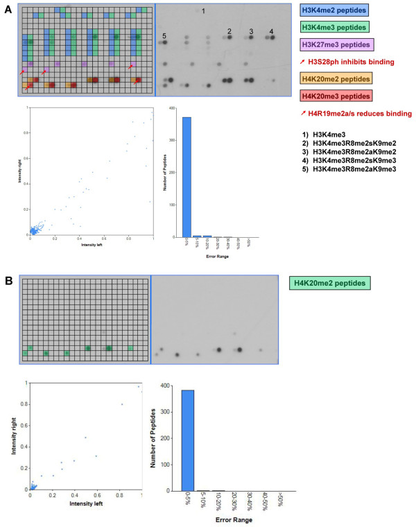
Background: Epigenetic reading domains are involved in the regulation of gene expression and chromatin state by interacting with histones in a post-translational modification specific manner. A detailed knowledge of the target modifications of reading domains, including enhancing and inhibiting secondary modifications, will lead to a better understanding of the biological signaling processes mediated by reading domains.

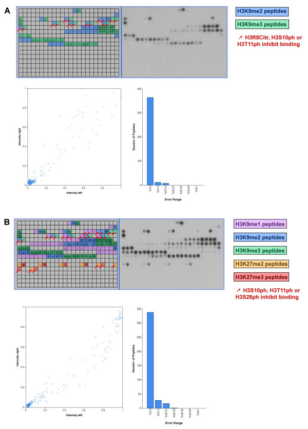
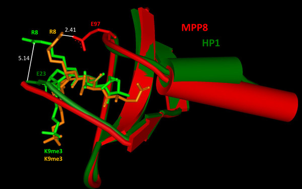
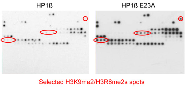
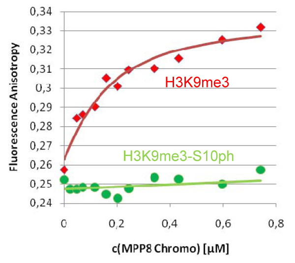
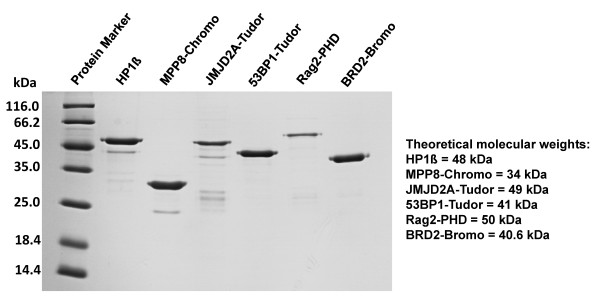
Results: We describe the application of Celluspots peptide arrays which contain 384 histone peptides carrying 59 post translational modifications in different combinations as an inexpensive, reliable and fast method for initial screening for specific interactions of reading domains with modified histone peptides. To validate the method, we tested the binding specificities of seven known epigenetic reading domains on Celluspots peptide arrays, viz. the HP1ß and MPP8 Chromo domains, JMJD2A and 53BP1 Tudor domains, Dnmt3a PWWP domain, Rag2 PHD domain and BRD2 Bromo domain. In general, the binding results agreed with literature data with respect to the primary specificity of the reading domains, but in almost all cases we obtained additional new information concerning the influence of secondary modifications surrounding the target modification.

Conclusions: We conclude that Celluspots peptide arrays are powerful screening tools for studying the specificity of putative reading domains binding to modified histone peptides.


Figures

Figure 1
Figure 1
Analysis of the binding specificity of Chromo domains on Celluspots peptide arrays. A) Binding analysis of HP1ß. The upper part of each array analysis shows the image of the array. Peptide spots are annotated on the left copy of the duplicates. The color code is described on the right side of the image. The red arrows indicate unbound peptide spots carrying the target modifications and secondary inhibiting modifications, which are specified on the right side of the image. In the lower part of the figure, a scatter plot of the binding intensities to corresponding peptides on both identical copies of the array and a bar diagram indicating the range of deviations is shown. B) Binding analysis of the MPP8 Chromo domain. For a description of the figure see legend of panel A.
Figure 2
Figure 2
Superposition of the HP1ß (pdb entry 1KNE) (dark green) and MPP8 Chromo domains (pdb entry 3QO2) (red) in complex with H3K9me3 peptides (light green for HP1 and orange for MPP8). 232 backbone atoms of both domains were superimposed with a root mean square of 0.92 Å using Deep View Swiss PDB viewer 3.7. The distance between the closest atoms of E23 of HP1 and R8 of the peptide is 5.14 Å. In MPP8, E97 approaches R8 more closely, with a distance of only 2.41 Å between the closest atoms.
Figure 3
Figure 3
Binding of wild type HP1ß and its E23A variant to Celluspots arrays showing similar binding specificity. Highlighted are some of the spots which carry H3K9me2 and H3R8me2s, some of them together with other modifications. The E23A variant binds better to these spots indicating that the negative effect of H3R8me2s on binding of H3K9me2 is reduced by the E23A mutation.
Figure 4
Figure 4
Binding of the MPP8 Chromo domain to H3K9me3 (red) and H3K9me3-S10ph peptides (green) analyzed in solution by fluorescence depolarization. Binding constants were determined by fitting of the data to a binary binding equilibrium to be 0.12 μM for H3K9me3 similarly as observed recently by isothermal calorimetry [13] and > 50 μM for H3K9me3-S10ph.
Figure 5
Figure 5
Analysis of the binding specificity of the JMJD2A double Tudor (A) and 53BP1 tandem Tudor (B) domains on Celluspots peptide arrays. For a description of the figure see the legend of figure 1A.
Figure 6
Figure 6
Analysis of the binding specificity of the Dnmt3a PWWP domain (A), the Rag2 PHD finger (B) and the BRD2 second Bromo domain (C) on Celluspots peptide arrays. For a description of the figure see legend of figure 1A.
Figure 7
Figure 7
Interaction between JMJD2A double Tudor domain and R19 in H4K20me3 peptide. The backbone of JMJD2A double Tudor domain (pdb entry 2QQS) is shown in orange. Amino acid side chains of the Tudor domains either interacting with H4K20me3 or H4R19 are shown in red. The H4-peptide backbone is shown in a medium blue with the side chains of K20me3 and R19 highlighted in a dark blue. The side chains of F932, D934, Y973 and W967 form an aromatic cage around the trimethylated lysine 20, while the side chains of F937 and D939 interact with unmodified R19. The distances of the side chains atoms of D939 and F937 to R19 are indicated.
Figure 8
Figure 8
Coomassie stained SDS gel showing the purified proteins used for peptide binding experiments. Loading was not adjusted to protein concentrations. The purification of the Dnmt3a PWWP domain is shown in Dhayalan et al. (2010) [19].

References

    1. Kouzarides T. Chromatin modifications and their function. Cell. 2007;128(4):693–705. doi: 10.1016/j.cell.2007.02.005. - DOI - PubMed
    1. Lee JS, Smith E, Shilatifard A. The language of histone crosstalk. Cell. 2010;142(5):682–685. doi: 10.1016/j.cell.2010.08.011. - DOI - PMC - PubMed
    1. Berger SL. The complex language of chromatin regulation during transcription. Nature. 2007;447(7143):407–412. doi: 10.1038/nature05915. - DOI - PubMed
    1. Sims RJ, Reinberg D. Is there a code embedded in proteins that is based on post-translational modifications? Nat Rev Mol Cell Biol. 2008;9(10):815–820. doi: 10.1038/nrm2502. - DOI - PubMed
    1. Taverna SD, Li H, Ruthenburg AJ, Allis CD, Patel DJ. How chromatin-binding modules interpret histone modifications: lessons from professional pocket pickers. Nat Struct Mol Biol. 2007;14(11):1025–1040. doi: 10.1038/nsmb1338. - DOI - PMC - PubMed

Publication types

MeSH terms

Substances

LinkOut - more resources