. 2011 Oct 25;419(2):97-106.
doi: 10.1016/j.virol.2011.08.005. Epub 2011 Aug 31.

Parainfluenza virus 5-based vaccine vectors expressing vaccinia virus (VACV) antigens provide long-term protection in mice from lethal intranasal VACV challenge

Affiliations

Parainfluenza virus 5-based vaccine vectors expressing vaccinia virus (VACV) antigens provide long-term protection in mice from lethal intranasal VACV challenge

Kimberly M Clark et al. Virology. .

Abstract

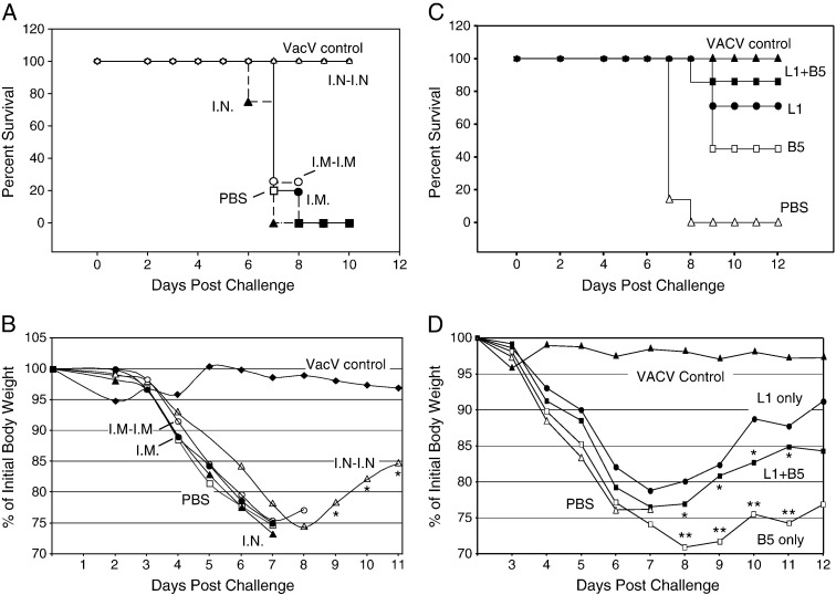
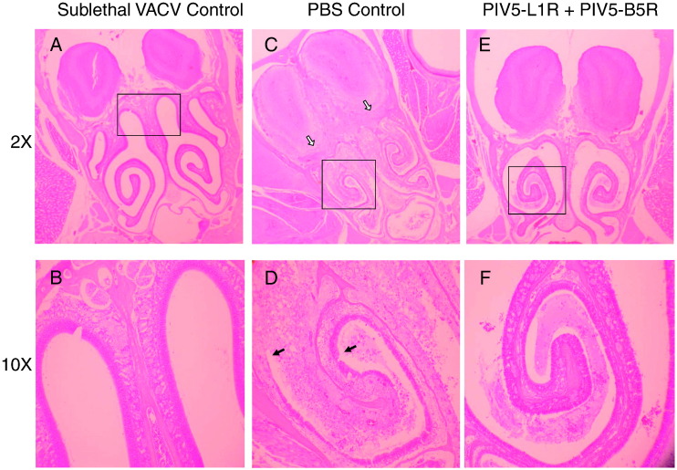
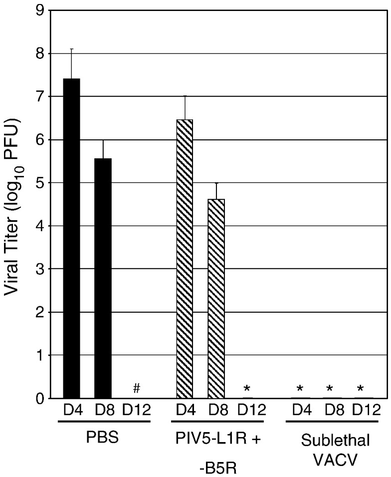
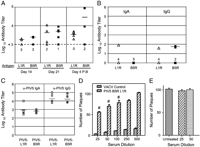
To test the potential for parainfluenza virus 5 (PIV5)-based vectors to provide protection from vaccinia virus (VACV) infection, PIV5 was engineered to express secreted VACV L1R and B5R proteins, two important antigens for neutralization of intracellular mature (IMV) and extracellular enveloped (EEV) virions, respectively. Protection of mice from lethal intranasal VACV challenge required intranasal immunization with PIV5-L1R/B5R in a prime-boost protocol, and correlated with low VACV-induced pathology in the respiratory tract and anti-VACV neutralizing antibody. Mice immunized with PIV5-L1R/B5R showed some disease symptoms following VACV challenge such as loss of weight and hunching, but these symptoms were delayed and less severe than with unimmunized control mice. While immunization with PIV5 expressing B5R alone conferred at least some protection, the most effective immunization included the PIV5 vector expressing L1R alone or in combination with PIV5-B5R. PIV5-L1R/B5R vectors elicited protection from VACV challenge even when CD8+ cells were depleted, but not in the case of mice that were defective in B cell production. Mice were protected from VACV challenge out to at least 1.5 years after immunization with PIV5-L1R/B5R vectors, and showed significant levels of anti-VACV neutralizing antibodies. These results demonstrate the potential for PIV5-based vectors to provide long lasting protection against complex human respiratory pathogens such as VACV, but also highlight the need to understand mechanisms for the generation of strong immune responses against poorly immunogenic viral proteins.


Figures

Fig. 1
Fig. 1
Expression of L1R and B5R from recombinant PIV5 vectors. A) Schematic diagram of PIV5 viruses used in this study. The genome structure of PIV5 is shown schematically as negative sense RNA with the insertion site for L1R and B5R genes between HN and L. Black bar denotes HA tag added to the L1R and B5R open reading frames. le, leader; tr, trailer. B) Protein expression. A549 cells were infected at an MOI of 25 with PIV5 vectors expressing TK, L1R or B5R. At the indicated time pi, media and cell lysates were analyzed by western blotting for HA-tagged proteins or for NP. 5× denotes the L1R portion of the gel exposed 5 times longer.
Fig. 2
Fig. 2
Mice vaccinated with PIV5-L1R and PIV5-B5R are protected from lethal VACV challenge. Panels A and B) Groups of 5 mice each were immunized with PBS as a control or a combination of 106 PFU of PIV5-L1R plus PIV5-B5R through either the I.N or I.M route. On day 28, PIV5-vaccinated mice were boosted with either PBS as a control (I.N or I.M only) or with the same dose of PIV5 vectors by the I.N. or I.M. route (I.N.–I.N. or I.M.–I.M groups). Control groups received either a sublethal level of VACV I.N. (closed diamond, VACV control) or prime-boost with PBS (open squares). Twenty one days after the boost, mice were challenged with VACV equivalent to 20 MTD50 by I.N. administration. Mice were monitored daily to determine survival (panel A) and weight loss (panel B) as a percent of initial body weight. Mice were euthanized at an experimental end point when they had a loss of 30% of initial weight or development of severe disease characteristics that were indicative of terminal stages of disease. For panel A, p < 0.003 for comparison of I.N.–I.N. to I.N. only and also for I.N.–I.N. compared to I.M.–I.M. For panel B, * denotes p < 0.001 for comparison of I.N.–I.N. to VACV control. Panels C and D) Groups of 7 mice each were administered prime and boost I.N. doses of PBS (open triangle), a sublethal dose of VACV (VACV control, closed triangle), or 106 PFU of PIV5-L1R and PIV5-B5R alone (open square and closed circle) or in combination (closed square) on the same days described for panels A and B. Mice were then challenged with VACV and then monitored daily as in panel A. For panel C, p < 0.02 for comparison of VACV, L1 + B5, L1, and B5 groups compared to PBS control. For panel D, **; p < 0.005; *, p < 0.5 when comparing weight loss between animals immunized with PIV5-B5R alone to PIV5-L1R alone or PIV5-L1R plus PIV5-B5R, respectively.
Fig. 3
Fig. 3
Pathology in nasal tissue of vaccinated mice after lethal I.N. VACV challenge. Mice were vaccinated I.N. with PBS, a sublethal dose of VACV, or prime and boost with 106 PFU of both PIV5-L1R and PIV5-B5R as described in the legend to Fig. 2. Twenty one days after the boost, mice were challenged I.N. with 20 MTD50 of VACV. Seven d after challenge, tissues were evaluated histologically. Pictures represent H&E staining of a cross section of nasal cavities and olfactory lobes of the brain. Boxes in 2× pictures outline the area shown in higher magnification in the 10× panels. Panels C and D from the PBS-treated control mice show necrosis of the nasal epithelium with intense inflammation which extends into the brain through the cribiform plate. White arrows (2×) indicate the disruption of integrity of cribiform plate into the adjacent olfactory lobes and adjacent inflammation. The black arrows (10×) show areas of nasal epithelial ulceration. Panels E and F from mice immunized with the PIV5-L1R and PIV5-B5R show minimal fibrinous inflammatory exudate in the nasal cavity and minimal tissue disruption. Panels A and B from the sublethal VACV control animal are within normal limits.
Fig. 4
Fig. 4
VACV load in lungs of mice vaccinated with PIV5-L1R and PIV5-B5R vectors. Groups of 7 mice were vaccinated I.N. with PBS (black bars), a sublethal dose of VACV, or prime and boost with 106 PFU of both PIV5-L1R and PIV5-B5R (hatched bars) as described in the legend to Fig. 2. Twenty one days after the boost, mice were challenged I.N. with 20 MTD50 of VACV and levels of VACV in lungs were determined by plaque assay. Standard deviations are indicated by the error bars. *, no detectable virus; #, mice succumbed to infection.
Fig. 5
Fig. 5
Protection from lethal VACV challenge elicited by PIV5-L1R and PIV5-B5R vectors requires B cells but not CD8+ cells. A) Groups of 7 mice each were immunized by prime-boost I.N. with PBS (closed triangles), a sublethal dose of VACV (closed squares), or 106 PFU of PIV5-L1R and PIV5-B5R (open circles) as described in the legend to Fig. 2. CD8-positive cells were depleted by daily administration of anti-CD8 antibody for 4 day as described in Materials and methods. Mice were then challenged with 20 MTD50 of VACV by I.N. administration, and then monitored daily to determine survival. * and ** denote p < 0.001 and p < 0.04 for comparison of PIV5 vaccinated and VV control vaccinated animals to PBS vaccinated animals, respectively. B) Jh B cell-deficient mice were vaccinated I.N. with as described in the legend to Fig. 2. Twenty one days after the boost, mice were challenged I.N. with 20 MTD50 of VACV, and then monitored daily to determine survival. ^ denotes p < 0.022 for comparison of PIV5 vaccinated to VV control vaccinated animals.
Fig. 6
Fig. 6
PIV5-L1 and PIV5-B5 elicit neutralizing anti-L1 and anti-B5 antibodies. A–C) Groups of 5 mice were vaccinated I.N. with 106 PFU of either PIV5-L1 or PIV5-B5R and serum was collected at days 14 and 21. Mice were boosted with an equivalent dose of the same virus and serum and BAL was collected on days 4 and 10 post boost, respectively. Samples were analyzed by ELISA for anti-L1R (open triangles) or anti-B5R (closed boxes) IgG in serum (panel A). Alternatively, BAL was analyzed by ELISA for levels of IgA and IgG specific for L1R or B5R (panel B) or PIV5 (panel C). In panel A, the dotted line at 103 indicates background level for nonspecific reaction. Numbers below the symbols indicate the number of animals that did not sero-covert. Mean titers for a given set of animals are indicated by a horizontal bar. For panels A and B, titers were not statistically above that of negative controls. D and E) Sera from mice immunized with a combination of PIV5-L1R/B5R vectors (panel D) or from control mice immunized with PBS (panel E) were analyzed in an in vitro neutralization assay with 100 PFU of IMV VACV as described in Materials and methods. Serum from mice given a sublethal dose of VACV was analyzed as a positive control. Results are the mean of sera from 5 mice with standard deviation shown by the bar. #, p < 0.01 when comparing between the 100 PFU of VACV used as starting infectivity versus the PFU remaining when treated with serum from immunized mice.
Fig. 7
Fig. 7
Mice immunized with PIV5-L1R and PIV5-B5R vectors show low neutralizing antibody against the EEV form of VACV. Groups of 5 mice each were immunized by prime-boost I.N. with PBS, a sublethal dose of VACV, PIV5 lacking an inserted gene or a combination of 106 PFU of both PIV5-L1R plus PIV5-B5R as described in the legend to Fig. 2. Sera collected at 14 day post boost were diluted 1:20, 1:40 or 1:80 and then tested in a neutralization assay with 100 PFU of the EEV form of VACV. *, **, *** denote p < 0.14, p < 0.0012 and p < 0.0001 when compared to PBS control serum at 1:20 dilution.
Fig. 8
Fig. 8
Antibody responses and protection from lethal VACV challenge in mice 1.5 years after vaccination with PIV5-L1R and PIV5-B5R vectors. Groups of 5 mice each were immunized by prime-boost I.N. with PBS, a sublethal dose of VACV or a combination of 106 PFU of both PIV5-L1R plus PIV5-B5R as described in the legend to Fig. 2. After 1.5 years, mice were challenged with VACV equivalent to 20 MTD50 by I.N. administration. Mice were monitored daily to determine survival (panel A) and weight loss (panel B) as a percent of initial body weight. In panel A, * denotes an animal that succumbed to an infection unrelated to VV challenge. # denotes p < 0.003 in comparison of immunized mice to PBS control. In panel B, * denotes p < 0.01, when comparing animals immunized with PIV5-L1R/B5R to VACV control. Panels C and D) Serum from mice vaccinated with PIV5-L1R and -B5R (panel C) or with sublethal VACV (panel D) were tested in an in vitro neutralization assay with 100 PFU of VACV as described in Materials and methods. Results are the mean values from 5 mice with standard deviations indicated by the error bars. ^, p < 0.001 when comparing immune serum to PBS control.

Similar articles

Cited by

References

    1. Aldaz-Carroll L., Whitbeck J.C., Ponce de Leon M., Lou H., Hirao L., Isaacs S.N., Moss B., Eisenberg R.J., Cohen G.H. Epitope-mapping studies define two major neutralization sites on the vaccinia virus extracellular enveloped virus glycoprotein B5R. J. Virol. 2005;79:6260–6271. - PMC - PubMed
    1. Aldaz-Carroll L., Whitbeck J.C., Ponce de Leon M., Lou H., Pannell L.K., Lebowitz J., Fogg C., White C.L., Moss B., Eisenberg R.J., Cohen G.H. Physical and immunological characterization of a recombinant secreted form of the membrane protein encoded by the vaccinia virus L1R gene. Virology. 2005;341:59–71. - PubMed
    1. Arimilli S., Johnson J.B., Clark K.M., Graff A.H., Alexander-Miller M.A., Mizel S.B., Parks G.D. Engineered expression of the TLR5 ligand flagellin enhances paramyxovirus activation of human dendritic cell function. J. Virol. 2008;82:10975–10985. - PMC - PubMed
    1. Bell E., Shamim M., Whitbeck J.C., Sfyroera G., Lambris J.D., Isaacs S.N. Antibodies against the EEV B5R protein are mainly responsible for EEV neutralization capacity of vaccinia virus immune globulin. Virology. 2004;325:425–431. - PubMed
    1. Belyakov I.M., Earl P., Dzutsev A., Kuznetsov V.A., Lemon M., Wyatt L.S., Snyder J.T., Ahlers J.D., Franchini G., Moss B., Berzofsky J.A. Shared modes of protection against poxvirus infection by attenuated and conventional smallpox vaccine viruses. Proc. Natl. Acad. Sci. U.S.A. 2005;100:9458–9463. - PMC - PubMed

Publication types

MeSH terms