

Monitoring tumor motion by real time 2D/3D registration during radiotherapy
- PMID: 21885144
- PMCID: PMC3276833
- DOI: 10.1016/j.radonc.2011.07.031
Monitoring tumor motion by real time 2D/3D registration during radiotherapy
Abstract
Background and purpose: In this paper, we investigate the possibility to use X-ray based real time 2D/3D registration for non-invasive tumor motion monitoring during radiotherapy.
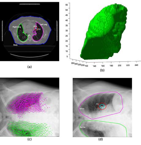
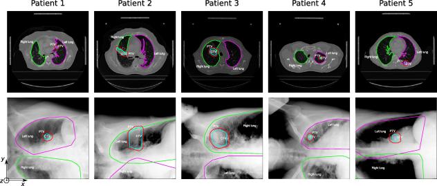
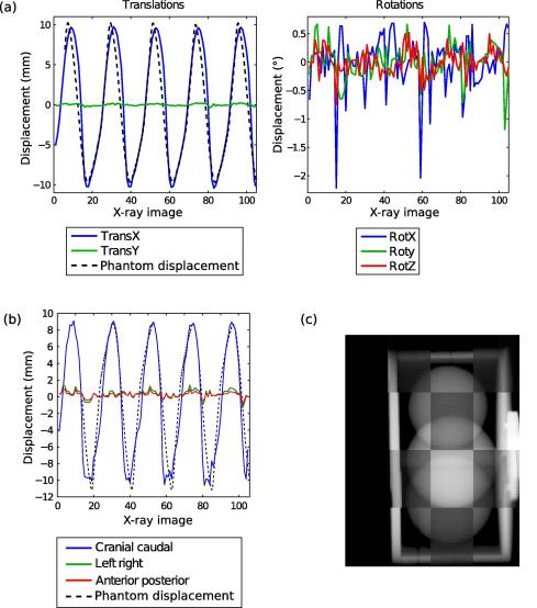
Materials and methods: The 2D/3D registration scheme is implemented using general purpose computation on graphics hardware (GPGPU) programming techniques and several algorithmic refinements in the registration process. Validation is conducted off-line using a phantom and five clinical patient data sets. The registration is performed on a region of interest (ROI) centered around the planned target volume (PTV).
Results: The phantom motion is measured with an rms error of 2.56 mm. For the patient data sets, a sinusoidal movement that clearly correlates to the breathing cycle is shown. Videos show a good match between X-ray and digitally reconstructed radiographs (DRR) displacement. Mean registration time is 0.5 s.
Conclusions: We have demonstrated that real-time organ motion monitoring using image based markerless registration is feasible.
Copyright © 2011 Elsevier Ireland Ltd. All rights reserved.
Figures

Similar articles
-
A novel bone suppression algorithm in intensity-based 2D/3D image registration for real-time tumor motion monitoring: Development and phantom-based validation.Med Phys. 2022 Aug;49(8):5182-5194. doi: 10.1002/mp.15716. Epub 2022 Jun 6. Med Phys. 2022. PMID: 35598307 Free PMC article.
-
High-performance GPU-based rendering for real-time, rigid 2D/3D-image registration and motion prediction in radiation oncology.Z Med Phys. 2012 Feb;22(1):13-20. doi: 10.1016/j.zemedi.2011.06.002. Epub 2011 Jul 22. Z Med Phys. 2012. PMID: 21782399 Free PMC article.
-
Investigation of simple IMRT delivery techniques for non-small cell lung cancer patients with respiratory motion using 4DCT.Med Dosim. 2009 Summer;34(2):158-69. doi: 10.1016/j.meddos.2008.07.001. Epub 2008 Aug 12. Med Dosim. 2009. PMID: 19410146
-
Evaluation of image guided motion management methods in lung cancer radiotherapy.Med Phys. 2014 Mar;41(3):031911. doi: 10.1118/1.4866220. Med Phys. 2014. PMID: 24593729
-
Nonrigid registration method to assess reproducibility of breath-holding with ABC in lung cancer.Int J Radiat Oncol Biol Phys. 2005 Feb 1;61(2):594-607. doi: 10.1016/j.ijrobp.2004.08.007. Int J Radiat Oncol Biol Phys. 2005. PMID: 15667982 Clinical Trial.
Cited by
-
[Image-guided radiation therapy].Radiologe. 2012 Mar;52(3):213-21. doi: 10.1007/s00117-011-2192-0. Radiologe. 2012. PMID: 22374083 German.
-
Artificial intelligence in image-guided radiotherapy: a review of treatment target localization.Quant Imaging Med Surg. 2021 Dec;11(12):4881-4894. doi: 10.21037/qims-21-199. Quant Imaging Med Surg. 2021. PMID: 34888196 Free PMC article. Review.
-
A novel bone suppression algorithm in intensity-based 2D/3D image registration for real-time tumor motion monitoring: Development and phantom-based validation.Med Phys. 2022 Aug;49(8):5182-5194. doi: 10.1002/mp.15716. Epub 2022 Jun 6. Med Phys. 2022. PMID: 35598307 Free PMC article.
-
2D/4D marker-free tumor tracking using 4D CBCT as the reference image.Phys Med Biol. 2014 May 7;59(9):2219-33. doi: 10.1088/0031-9155/59/9/2219. Epub 2014 Apr 8. Phys Med Biol. 2014. PMID: 24710793 Free PMC article.
-
Markerless tumor tracking using fast-kV switching dual-energy fluoroscopy on a benchtop system.Med Phys. 2019 Jul;46(7):3235-3244. doi: 10.1002/mp.13573. Epub 2019 Jun 1. Med Phys. 2019. PMID: 31059124 Free PMC article.
References
-
- Guckenberger M., Krieger T., Richter A., Baier K., Wilbert J., Sweeney R.A. Potential of image-guidance, gating and real-time tracking to improve accuracy in pulmonary stereotactic body radiotherapy. Radiother Oncol. 2009;91:288–295. - PubMed
-
- Han K., Cheung P., Basran P.S., Poon I., Yeung L., Lochray F. A comparison of two immobilization systems for stereotactic body radiation therapy of lung tumors. Radiother Oncol. 2010;95:103–108. - PubMed
-
- Korreman S., Rasch C., McNair H., Verellen D., Oelfke U., Maingon P. The European Society of Therapeutic Radiology and Oncology-European Institute of Radiotherapy (ESTRO-EIR) report on 3D CT-based in-room image guidance systems: a practical and technical review and guide. Radiother Oncol. 2010;94:129–144. - PubMed
-
- van Herk M. Errors and margins in radiotherapy. Semin Radiat Oncol. 2004;14:52–64. - PubMed
-
- Schweikard A., Glosser G., Bodduluri M., Murphy M.J., Adler J.R. Robotic motion compensation for respiratory movement during radiosurgery. Comput Aided Surg. 2000;5:263–277. - PubMed
Publication types
MeSH terms
Grants and funding
LinkOut - more resources
Full Text Sources
Other Literature Sources
Medical
