. 2011 Oct 28;286(43):37335-46.
doi: 10.1074/jbc.M111.256156. Epub 2011 Sep 1.

Lung tumor-associated osteoblast-derived bone morphogenetic protein-2 increased epithelial-to-mesenchymal transition of cancer by Runx2/Snail signaling pathway

Affiliations

Lung tumor-associated osteoblast-derived bone morphogenetic protein-2 increased epithelial-to-mesenchymal transition of cancer by Runx2/Snail signaling pathway

Ya-Ling Hsu et al. J Biol Chem. .

Abstract

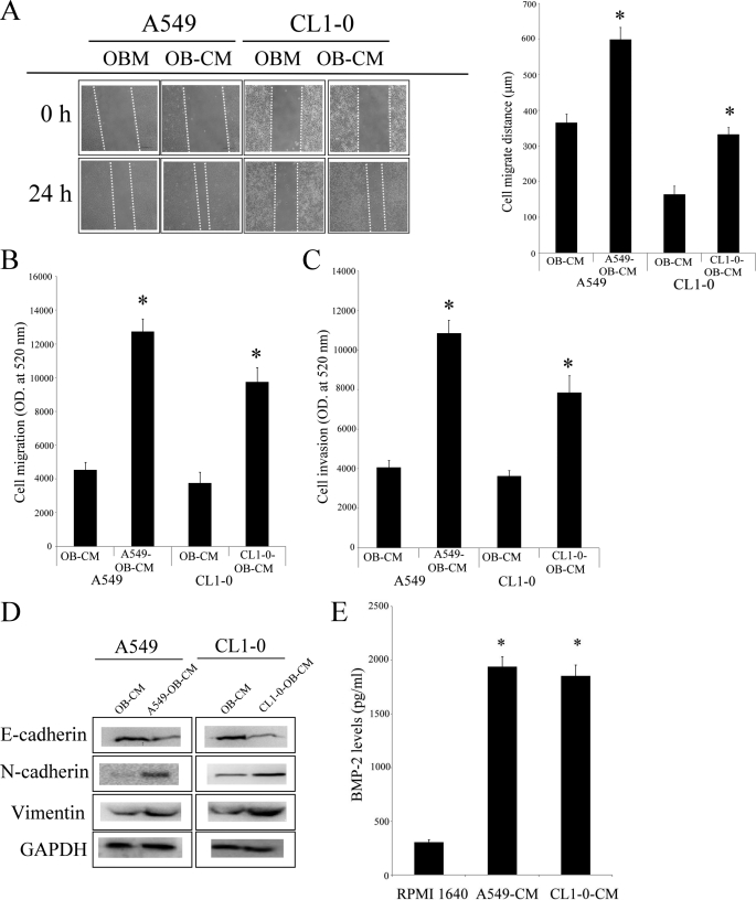
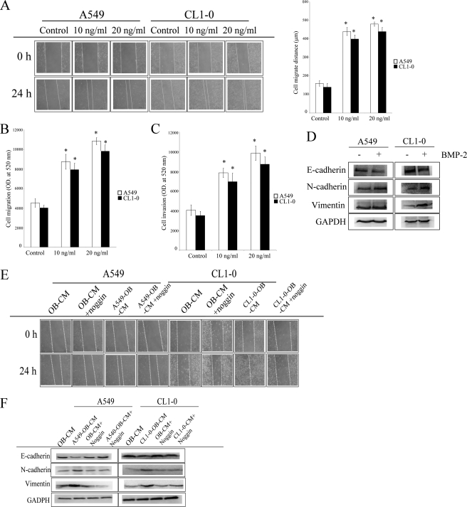
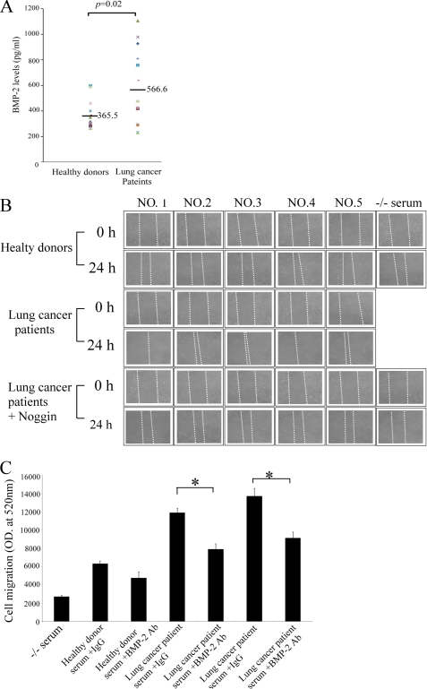
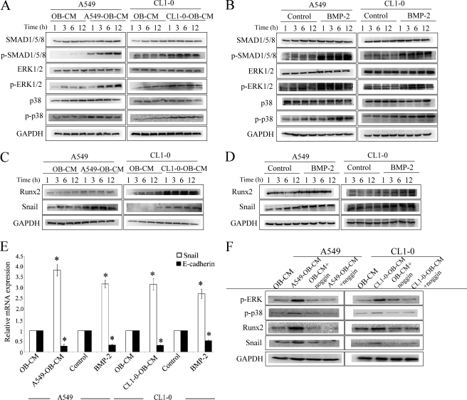
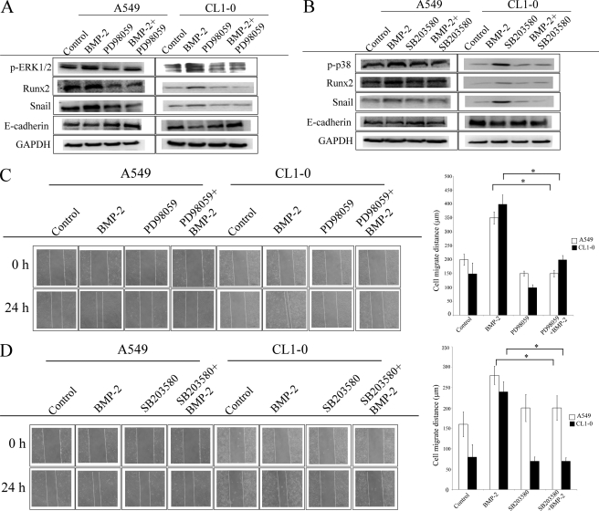
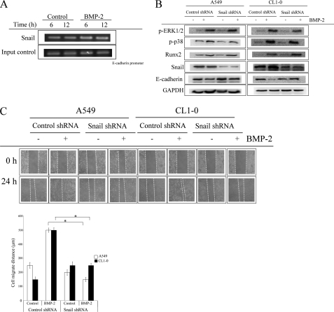
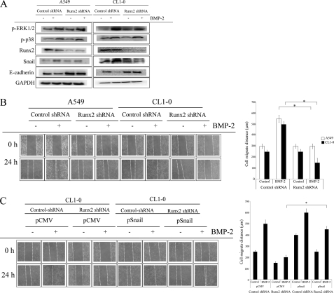
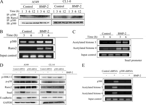
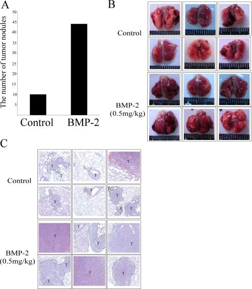
Bone is a frequent target of lung cancer metastasis and is associated with significant morbidity and a dismal prognosis. Interaction between cancer cells and the bone microenvironment causes a vicious cycle of tumor progression and bone destruction. This study analyzed the soluble factors secreted by lung tumor-associated osteoblast (TAOB), which are responsible for increasing cancer progression. The addition of bone morphogenetic protein-2 (BMP-2), present in large amounts in TAOB conditioned medium (TAOB-CM) and lung cancer patient sera, mimicked the inductive effect of TAOB-CM on lung cancer migration, invasion, and epithelial-to-mesenchymal transition. In contrast, inhibition of BMP by noggin decreases the inductive properties of TAOB-CM and lung cancer patient sera on cancer progression. Induction of lung cancer migration by BMP-2 is associated with increased ERK and p38 activation and the up-regulation of Runx2 and Snail. Blocking ERK and p38 by a specific inhibitor significantly decreases cancer cell migration by inhibiting Runx2 up-regulation and subsequently attenuating the expression of Snail. Enhancement of Runx2 facilitates Rux2 to recruit p300, which in turn enhances histone acetylation, increases Snail expression, and decreases E-cadherin. Furthermore, inhibiting Runx2 by siRNA also suppresses BMP-2-induced Snail up-regulation and cell migration. Our findings provide novel evidence that inhibition of BMP-2 or BMP-2-mediated MAPK/Runx2/Snail signaling is an attractive therapeutic target for osteolytic bone metastases in lung cancer patients.


Figures

FIGURE 1.
FIGURE 1.
TAOB-CMs increases lung cancer migration and EMT. TAOB-CMs enhanced the migratory ability of A549 and CL1–0 lung cancer cells. A and B, TAOB-CMs enhanced cell migratory ability, as determined by scratch wound healing assay (A) and Transwell system (B). C, TAOB-CMs enhanced cell invasion ability. D, TAOB-CMs caused EMT in cancer cells. E, the expression of BMP-2 in OB-CM, For A, the migration ability of lung cancer cells was assessed by wound healing assay. OB-CM (control group) and TAOB-CMs (20%) act as a chemoattractant of cancer migration. Quantification of cell migration was carried out by measuring the distance between the migratory fronts of cells in four random selected microscopic fields for each condition and time point. The degree of cell movement is expressed as the percentage of wound closure as compared with the zero time point. For B and C, the invasiveness and migration ability of A549 and CL1–0 cells were quantified by QCMTM 24-well cell migration and invasion assay. The cells were seeded in the upper inset, and the OB-CM (control group) and TAOB-CMs (20%) acted as the chemoattractant for cancer migration and invasion. For D, A549 and CL1–0 cells were treated with TAOB-CMs (20%) for 24 h, and then the expression of various proteins was assessed by immunoblot assay. For E, primary osteoblasts were treated RPMI 1640 (20%), A549-CM (20%), and CL1–0-CM for 24 h. The BMP-2 levels were assessed by BMP-2 ELISA kits. Each value is the mean ± S.D. of three independent experiments. The asterisk indicates a significant difference between control and test groups, as analyzed by Dunnett's test (p < 0.05).
FIGURE 2.
FIGURE 2.
BMP-2 is involved in TAOB-CM-mediated enhancement of migration and EMT in lung cancer. A and B, BMP-2 increased migratory ability, as determined by scratch wound healing assay (A) and Transwell system (B). C and D, BMP-2 increased the invasion ability (C) and EMT (D) of A549 and CL1–0 cells. E and F, noggin decreased TAOB-CM-mediated cell migration (E) and EMT (F). The migration ability of lung cancer cells was assessed by wound healing assay, in accord with the description under “Experimental Procedures.” BMP-2 (20 ng/ml for EMT assay) acts as the chemoattractant for cancer migration. For E and F, A549 and CL1–0 cells were pretreated with or without noggin for 1 h, and then OB-CM and TAOB-CMs were added for another 24 h. Cell migration was assessed by wound healing assay, and the expression of various proteins was determined by immunoblot assay. Each value is the mean ± S.D. of three independent experiments. The asterisk indicates a significant difference between control and test groups, as analyzed by Dunnett's test (p < 0.05).
FIGURE 3.
FIGURE 3.
Sera from lung cancer patients increase lung cancer migration. A, the levels of BMP-2 in lung cancer patient sera. B, lung cancer sera enhance the migratory ability of lung cancer cells. C, depletion of BMP-2 decreased lung cancer patient serum-mediated cell migration. The levels of BMP-2 were assessed by ELISA. Horizontal bars represent means. The cells were treated with or without noggin for 1 h, and then culture medium containing healthy donor sera (15%) or lung cancer patient sera (15%) was added for another 24 h. Cell migration was assessed by wound healing assay. For C, BMP-2 depleted from lung cancer patient serum was performed using anti-BMP-2 and antibodies (4 μg/ml) and Sepharose A/G beads, following regular immunoprecipitation techniques. The migration ability of A549 and CL1–0 cells were quantified by QCMTM 24-well cell migration assay kit. Each value is the mean ± S.D. of three independent experiments. The asterisk indicates a significant difference between control and test groups, as analyzed by Dunnett's test (p < 0.05).
FIGURE 4.
FIGURE 4.
TAOB-CMs and BMP-2 increase the activation of MAPK and elevate the expression of Runx2 and Snail. A and B, TAOB-CMs (A) and BMP-2 (B) increase the phosphorylation of SMAD, ERK, and p38. C and D, TAOB-CMs (C) and BMP-2 (D) enhance the expression of Runx2 and Snail protein. Cells were treated with OB-CM (20%), TAOB-CMs (20%), or BMP-2 (20 ng/ml) for the indicated times. The expressions of various proteins were determined by immunoblot assay. E, TAOB-CMs and BMP-2 enhance the expression of Runx2 and Snail mRNA. The cells were treated with OB-CM (20%), TAOB-CMs (20%), or BMP-2 (20 ng/ml) for a specific time (3 h for snail and 12 h for E-cadherin). The expressions of mRNA were determined by quantitative PCR. F, noggin decreases TAOB-CM-mediated MAPK activation and Runx2 and Snail up-regulation. The cells were treated with OB-CM (20%), TAOB-CMs (20%), or BMP-2 (20 ng/ml) for the indicated times. The expressions of mRNA and various proteins were determined by quantitative PCR and immunoblot assay. For F, A549 and CL1–0 cells were pretreated with or without noggin for 1 h and then treated with BMP-2 (20 ng/ml) for 6 h. The expression of various proteins was then assessed by immunoblot assay. The data shown are representative of three independent experiments. Each value is the mean ± S.D. of three independent experiments. The asterisk indicates a significant difference between control and test groups, as analyzed by Dunnett's test (p < 0.05).
FIGURE 5.
FIGURE 5.
The role of ERK1/2 and p38 on Runx2 and Snail expression. A and B, ERK inhibitor (A) and p38 inhibitor (B) decrease BMP-2-mediated up-regulation of Runx2 and Snail as well as E-cadherin. C and D, ERK inhibitor (C) and p38 inhibitor (D) decrease BMP-2-mediated lung cancer cell migration. The cells were treated with PD98059 (ERK inhibitor, 10 μm) or SB203580 (p38 inhibitor, 10 μm) for 1 h, and then BMP-2 (20 ng/ml) was added for the specified times (cell migration, 24 h; Runx2 and Snail, 6 h; E-cadherin, 24 h). The expression of various proteins was then assessed by immunoblot assay. Cell migration was assessed by wound healing assay. The data shown are representative of three independent experiments. Each value is the mean ± S.D. of three independent experiments. The asterisk indicates a significant difference between control and test groups, as analyzed by Dunnett's test (p < 0.05).
FIGURE 6.
FIGURE 6.
Snail is involved in BMP-2-mediated cell migration and EMT. A, the binding of Snail on E-cadherin promoter. B and C, knockdown of Snail decreases BMP-2 mediated E-cadherin down-regulation (B) and cell migration (C). The cells were treated with BMP-2 (20 ng/ml) for the indicated times. The Snail binding on E-cadherin was determined by chromatin immunoprecipitation. The cells were transfected with pLKO-AS2 or pLKO-AS2-SNAIL1 shRNA. Stable clones were created by puromycin selection, and the efficacy of shRNA was assessed by RT-PCR. The cells were treated with BMP-2 (20 ng/ml) for the specified times (cell migration, 24 h; Runx2 and Snail, 6 h; E-cadherin, 24 h). Then the expression of various proteins was then assessed by immunoblot assay. Cell migration was assessed by wound healing assay. The data shown are representative of three independent experiments. Each value is the mean ± S.D. of three independent experiments. The asterisk indicates a significant difference between control and test groups, as analyzed by Dunnett's test (p < 0.05). The data shown are representative of three independent experiments.
FIGURE 7.
FIGURE 7.
Runx2 is the upstream regulatory factor of Snail. A and B, inhibition of Runx2 decreases BMP-2-mediated Snail up-regulation and E-cadherin down-regulation (A), as well as cell migration (B). Cells were transfected with pLKO-AS2 or pLKO-AS2-RUNX2 shRNA. Stable clones were created by puromycin selection, and the efficacy of shRNA was assessed by RT-PCR. Cells were treated with BMP-2 (20 ng/ml) for the specified times (cell migration, 24 h; Runx2 and Snail, 6 h; E-cadherin, 24 h). Then the expression of various proteins was then assessed by immunoblot assay. C, overexpression of Snail reversed the inhibitory effect of Runx2 shRNA on BMP-2-mediated cell migration. Runx2-transfected A549 and CL1–0 cells were transected with pCMV or pSnail plasmid, and stable clones were established by G418 and puromycin. The asterisk indicates a significant difference between control and test groups, as analyzed by Dunnett's test (p < 0.05). The data shown are representative of three independent experiments.
FIGURE 8.
FIGURE 8.
Runx2-p300 complex regulates the expression of Snail. A, the interaction of Runx2 with p300. B, the binding of p300 and Runx2 on Snail promoter. C, BMP-2 increased the acetylation of histone H3 and H4 on the p300-binding region of Snail promoter. D and E, inhibition of p300 prevents BMP-2-mediated Snail up-regulation (D) and histone acetylation on Snail promoter (E). The cells were treated with BMP-2 (20 ng/ml) for the indicated times. The interaction of Runx2 was assessed by immunoprecipitation (IP). The histone acetylation and p300/Runx2 binding on Snail were determined by chromatin immunoprecipitation. The cells were transfected with pLKO-AS2 or pLKO-AS2-p300. Stable clones were created by puromycin selection, and the efficacy of shRNA was assessed by RT-PCR. shRNA-transfected cells were treated with BMP-2 (20 ng/ml) for the specified times (Runx2 and Snail, 6 h; E-cadherin, 24 h). The expression of various proteins was then assessed by immunoblot (IB) assay. The data shown are representative of three independent experiments.
FIGURE 9.
FIGURE 9.
BMP-2 increased the metastasis of lung cancer cell in vivo. A, BMP-2 increased tumor nodules in lungs. B and C, BMP-2 increased lung metastasis as revealed by photographs (B) and hematoxylin and eosin staining (C). LLC cells were injected into mice via the tail vein. The mice were dosed every 3 days with intraperitoneal injections PBS (n = 8) or BMP-2 (0.5 mg/kg) (n = 7). After 10 days, nontumorous and tumorous regions of the lungs were harvested, cut, hematoxylin- and eosin-stained, and analyzed by microscopy. The number of tumor nodules was recorded for analysis of lung cancer incidence. The asterisk indicates a significant difference with the control, as analyzed by analysis of variance with Student's t test post hoc. *, p < 0.05.

Similar articles

Cited by

References

    1. Roodman G. D. (2004) N. Engl. J. Med. 350, 1655–1664 - PubMed
    1. Coleman R. E., Lipton A., Roodman G. D., Guise T. A., Boyce B. F., Brufsky A. M., Clézardin P., Croucher P. I., Gralow J. R., Hadji P., Holen I., Mundy G. R., Smith M. R., Suva L. J. (2010) Cancer Treat. Rev. 36, 615–620 - PMC - PubMed
    1. Rosen L. S., Gordon D., Tchekmedyian S., Yanagihara R., Hirsh V., Krzakowski M., Pawlicki M., de Souza P., Zheng M., Urbanowitz G., Reitsma D., Seaman J. J. (2003) J. Clin. Oncol. 21, 3150–3157 - PubMed
    1. Kvale P. A., Simoff M., Prakash U. B. (2003) Chest 123, (Suppl. 1) 284S–311S - PubMed
    1. Onishi T., Hayashi N., Theriault R. L., Hortobagyi G. N., Ueno N. T. (2010) Nat. Rev. Clin. Oncol. 7, 641–651 - PubMed

Publication types

MeSH terms