. 2011 Oct 28;286(43):37768-77.
doi: 10.1074/jbc.M111.281592. Epub 2011 Sep 1.

Oligomerization and pore formation by equinatoxin II inhibit endocytosis and lead to plasma membrane reorganization

Affiliations

Oligomerization and pore formation by equinatoxin II inhibit endocytosis and lead to plasma membrane reorganization

Ana J García-Sáez et al. J Biol Chem. .

Abstract

Pore-forming toxins have evolved to induce membrane injury by formation of pores in the target cell that alter ion homeostasis and lead to cell death. Many pore-forming toxins use cholesterol, sphingolipids, or other raft components as receptors. However, the role of plasma membrane organization for toxin action is not well understood. In this study, we have investigated cellular dynamics during the attack of equinatoxin II, a pore-forming toxin from the sea anemone Actinia equina, by combining time lapse three-dimensional live cell imaging, fluorescence recovery after photobleaching, FRET, and fluorescence cross-correlation spectroscopy. Our results show that membrane binding by equinatoxin II is accompanied by extensive plasma membrane reorganization into microscopic domains that resemble coalesced lipid rafts. Pore formation by the toxin induces Ca(2+) entry into the cytosol, which is accompanied by hydrolysis of phosphatidylinositol 4,5-bisphosphate, plasma membrane blebbing, actin cytoskeleton reorganization, and inhibition of endocytosis. We propose that plasma membrane reorganization into stabilized raft domains is part of the killing strategy of equinatoxin II.


Figures

FIGURE 1.
FIGURE 1.
EqtII binds rapidly to cells and induces plasma membrane blebbing. A, time series of EqtIIAl488 action on HEK273 cells followed simultaneously by differential interference contrast and epifluorescence. The differential interference contrast images in the top row show the appearance of plasma membrane blebs that grow over time. In the lower row, binding of EqtII-Al488 can be detected by epifluorescence a shortly after toxin addition. Scale bar, 20 μm. B, the size of EqtII-induced blebs is concentration-dependent. The images show the state of HEK273 cells after a 1-h addition of the indicated EqtII amounts. Scale bar, 20 μm. C, confocal three-dimensional projection of a HEK273 cell 5 min after the addition of EqtII-Al488 shows heterogeneous distribution of the protein on the cell surface. Scale bar, 10 μm. Toxin concentration in A and C was 5 μg/ml.
FIGURE 2.
FIGURE 2.
EqtII induces plasma membrane reorganization. A, time series of GPI-GFP redistribution in the membrane of COS-7 cells upon the addition of EqtII. The time points after toxin addition are indicated. The images are three-dimensional projections of confocal slices. Scale bar, 30 μm. B, FRAP experiments to characterize GPI-GFP dynamics in absence (upper panel) and presence (lower panel) of EqtII. Time points before and after bleaching and after the indicated recovery time are shown. The bleached areas correspond to the white circles. Scale bar, 20 μm. C, FRAP recovery curves of GPI-GFP fluorescence intensity in absence (blue line) and presence (red line) of EqtII. Curves are averages of five measurements, and original curves are shown in gray. Toxin concentration was 5 μg/ml.
FIGURE 3.
FIGURE 3.
EqtII colocalizes with raft markers on the plasma membrane. COS-7 cells transfected with the indicated plasma membrane proteins fused to fluorescent proteins were treated with EqtII-Atto655, and the distribution of the fluorescent markers was followed by three-dimensional time lapse microscopy (supplemental Movies S3–S7). The changes in protein distribution before (0 min) and 20 min after toxin addition are shown as Z projections. The colocalization between EqtII (red lines) and raft-associated GPI-GFP, Myr-Pal-YFP and LAT-GFP (blue lines) is shown in the column with intensity profiles corresponding to the white lines in the merged channels. In contrast, colocalization with non-raft-associated VSV-sp-GFP and PCX-GFP is not evident. Scale bar, 20 μm.
FIGURE 4.
FIGURE 4.
EqtII action induces Ca2+ entry, PI(4,5)P2 degradation, endocytosis inhibition and reorganization of the actin cytoskeleton. A, addition of EqtII induces an increase in intracellular Ca2+ in a few minutes (upper panel). This correlates with degradation of PI(4,5)P2 at the plasma membrane, as shown by the reorganization of PH(PLC)GFP fluorescence within the same time range. In contrast, Fluo4 signal does not show any increase, and PI(4,5)P2 signal is maintained at the plasma membrane when a buffer without Ca2+ is used (controls, 10 min). Scale bars, 25 μm. B, CTB-Al488 endocytosis is inhibited after the addition of EqtII. The first panel shows CTB-Al488 that has been endocytosed when it is added 2 h before EqtII. In the second and third panels, CTB-Al488 is not endocytosed when EqtII is added simultaneously or 5 min before CTB-Al488. The fourth panel shows small amounts of endocytosis when CBT-Al488 is added 5 min before EqtII. In all cases, the cells were analyzed after 1 h of incubation with EqtII at room temperature. Scale bar, 15 μm. C, quantification of endocytosed CTB-Al488 in the samples shown in B. The number of cells analyzed in each sample is shown. D, control of CBT-Al488 endocytosis after 1 h in the absence of EqtII. Scale bar, 15 μm. E, reorganization of actin cytoskeleton upon EqtII addition. Actin filaments stained with Lifeact-GFP underwent reorganization within the same time scales as the redistribution of membrane markers (Figs. 2 and 3). COS-7 cells were used in all experiments. Scale bar, 25 μm.
FIGURE 5.
FIGURE 5.
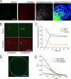
EqtII oligomerizes on the plasma membrane of target cells. A–C, EqtII oligomerization shown by FRET. EqtII-Al488 (donor) and EqtII-Al555 (acceptor) were added to COS-7 cells and incubated for 1 h at room temperature. In A, the fluorescence of the donor and acceptor channels is shown in the first and second panels, respectively. The third panel shows the calculated sensitized emission FRET for the same region, which is normalized to apparent FRET efficiency in the fourth panel. The color code goes from black for minimum gray values to blue, green, yellow, orange, red, and then white for maximum gray values. Scale bar, 15 μm. In B, EqtII oligomerization is shown by acceptor photobleaching FRET. The increase in the donor fluorescence in the region indicated with an arrow (upper panel) upon acceptor photobleaching within that region via high intensity illumination (lower panel) is a direct evidence of EqtII/EqtII interactions. C, time changes in the fluorescence detected in donor, acceptor, and FRET channels upon acceptor photobleaching. D and E, direct EqtII/EqtII interactions shown by two-color two-focus scanning FCS. COS-7 cells were treated with Eqt-Al488 and EqtII-Atto655 and incubated for 1 h at room temperature. FCS was measured on the membrane of cell blebs induced by the toxin. D, the two foci of scanning FCS are depicted as parallel lines on one of the blebs measured. E, auto- and cross-correlation fitted curves of EqtII-Al488 and EqtII-Atto655. The positive amplitude of the cross-correlation curve (blue line) indicates direct EqtII/EqtII interactions. Raw data are shown in black lines. Scale bars, 15 μm.
FIGURE 6.
FIGURE 6.
Model of EqtII-induced membrane reorganization during its toxic action. EqtII binds specifically to the SM exposed in the outer leaflet of the plasma membrane of cells and oligomerizes to form a pore. This allows calcium entry into the cytosol, which among other effects induces PI(4,5)P2 degradation, and endocytosis and cytoskeleton dynamics are inhibited. As a result, the alterations in membrane traffic affect the membrane repair machinery, and EqtII oligomers are not internalized, thus growing to immobile microscopic domains that accumulate other raftophilic proteins. In addition, membrane links to the cytoskeleton are disrupted and, together with osmotic imbalance, lead to the growth of stable blebs. Finally, EqtII-induced transformation into a dysfunctional plasma membrane, whose lateral organization and permeability are compromised, leads to cell death.

References

    1. Anderluh G., Lakey J. H. (2008) Trends Biochem. Sci. 33, 482–490 - PubMed
    1. Bischofberger M., Gonzalez M. R., van der Goot F. G. (2009) Curr. Opin. Cell Biol. 21, 589–595 - PubMed
    1. Kleanthous C. (2010) Nat. Rev. Microbiol 8, 843–848 - PubMed
    1. Belmonte G., Pederzolli C., Macek P., Menestrina G. (1993) J. Membr. Biol. 131, 11–22 - PubMed
    1. Mancheño J. M., Martín-Benito J., Gavilanes J. G., Vázquez L. (2006) Biophys. Chem. 119, 219–223 - PubMed

Publication types