. 2011 Oct;236(10):1211-7.
doi: 10.1258/ebm.2011.011053. Epub 2011 Sep 1.

Lentivirus-mediated expression of cDNA and shRNA slows degeneration in retinitis pigmentosa

Affiliations

Lentivirus-mediated expression of cDNA and shRNA slows degeneration in retinitis pigmentosa

Joaquin Tosi et al. Exp Biol Med (Maywood). 2011 Oct.

Abstract

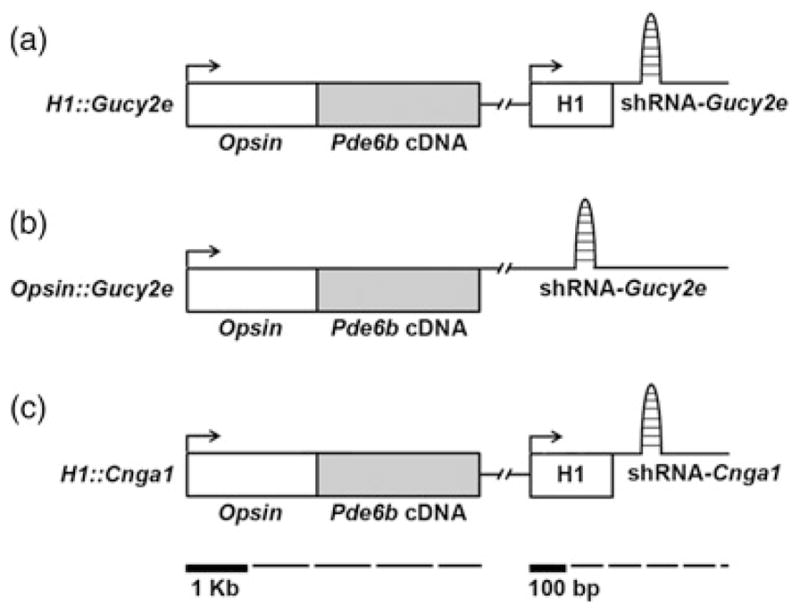
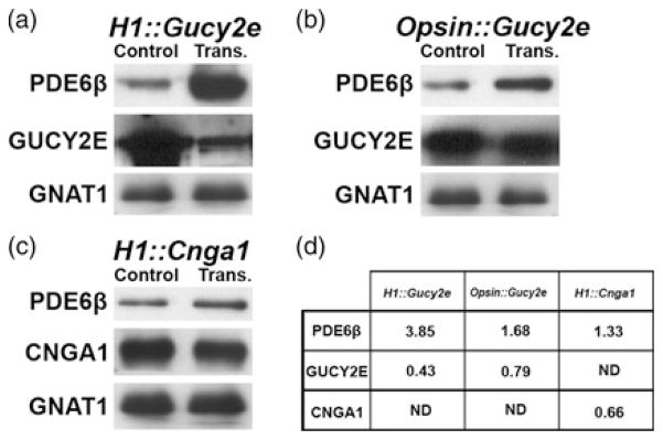
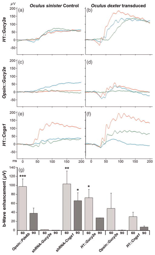
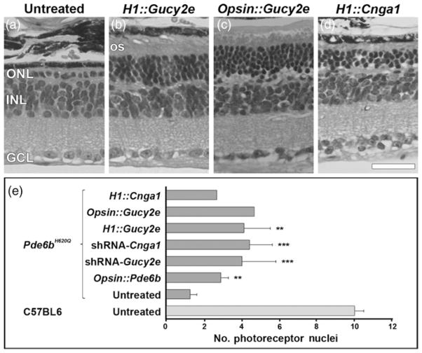
Mutations in Pde6b lead to high levels of signaling molecules cyclic guanosine monophosphate (cGMP) and Ca(2+), which ultimately result in photoreceptor cell death in certain forms of retinitis pigmentosa (RP). The level of cGMP, which is controlled by opposing activities of guanylate cyclase (GUCY) and photoreceptor phosphodiesterase-6 (PDE6), regulates the opening of cyclic nucleotide-gated ion channels [CNG] and thereby controls Ca(2+) influx into the outer segments. Using a lentiviral gene therapy approach, we have previously shown that degeneration can be temporarily slowed either by introducing wild-type PDE6β or knocking down expression of GUCY2E and CNGA1 in photoreceptors of Pde6b(H620Q), a mouse model for RP. Rescue was transient with either approach. Therefore, we tested a novel combination therapy using bipartite lentiviral vectors designed to both introduce wild-type PDE6β expression and knockdown GUCY2E or CNGA1. Immunoblot analysis shows simultaneous increases in PDE6β and decreases in GUCY2E or CNGA1 in retinas transduced by the vectors, indicating successful transduction. In Pde6b(H620Q) mutants, we observe rescue of photoreceptor function and an increase in photoreceptor rows as compared with untreated controls. However, no evidence of prolonged rescue beyond the limit of the previously tested single therapy was observed.


Figures

Figure 1
Figure 1
Schematic representation of the lentiviral vectors. The H1::Gucy2e (a) construct consists of a 2 kb mouse Opsin promoter, a Pde6b open reading frame (Pde6b cDNA) and the H1 promoter, which drives expression of the shRNA-Gucy2e. The Opsin::Gucy2e (b) consists of a 2 kb Opsin promoter, a Pde6b open reading frame and a shRNA-Gucy2e. The H1::Cnga1 lentiviral vector (c) was constructed using 2 kb of the mouse Opsin promoter, which directs expression of the Pde6b cDNA fragment, and the H1 promoter, which drives expression of the shRNA-Cnga1
Figure 2
Figure 2
Immunoblotting analysis of retinal lysates from C57BL/6J mice transduced with bipartite vectors. PDE6β was increased after transduction with either H1::Gucy2e, Opsin::Gucy2e or H1::Cnga1 (a–c). GUCY2E is depressed in retinas transduced with H1::Gucy2e (a) and Opsin::Gucy2e (b) compared with controls. CNGA1 expression is reduced in retinas transduced with H1::Cnga1 (c) compared with controls. GNAT1 was used as control for photoreceptor protein content and its expression was not reduced by any of the three viruses. Intensities were determined by densitometry to calculate an integral density value for each band. The values were normalized to total protein and GNAT1 expression as a percentage of GNAT1 signal in 20 μg of control lysate (d). Samples from each experimental group were pooled in order to minimize variation resulting from differences in subretinal surgeries; immunoblot analyses were carried out in duplicate (average values shown). ND, not determined; trans., transduced
Figure 3
Figure 3
Functional rescue due to bipartite vector transduction. Maximal dark-adapted electroretinograms (ERGs) were performed on both eyes (transduced and control) of Pde6bH620Q animals at approximately P60 and P90. In each ERG panel (a–f), the tracings represent different mice from the same treatment group. Compared with untreated controls (a, c, e), the H1::Gucy2e (b), Opsin::Gucy2e (d) and H1::Cnga1 (f) vectors improved visual function. Function was measured as both the photoreceptor-mediated a-wave and inner retina-mediated b-wave. ERG traces from three mice are shown for each virus. Enhancement of b-wave was calculated by comparing ERG values from transduced and untreated eyes. Results of the bipartite therapies described in this study (H1::Gucy2e, Opsin::Gucy2e and H1::Cnga1) were compared with previous results of the monopartite therapies (Opsin::Pde6b, shRNA-Gucy2e and shRNA-Cnga1), (g). Significant values: *P < 0.05, **P < 0.01, ***P < 0.001 (A color version of this figure is available in the online journal)
Figure 4
Figure 4
Photoreceptor cell rescue due to bipartite vector transduction. Hematoxylin–eosin labeling in Pde6bH620Q retinal sections at P60 stained the different layers of the retina: ONL, INL, GCL (a–d). In non-treated Pde6bH620Q retinas, the ONL contained only a single row of photoreceptors (a). Treated retinas with H1::Gucy (b), Opsin::Gucy (c) and H1::Cnga1 (d) showed a higher number of photoreceptor rows within the ONL. Outer segments (OS) were also preserved in transduced but not in untreated retinas. Photoreceptor cell rescue in Pde6bH620Q using bipartite therapies described in this study (H1::Gucy2e, Opsin::Gucy2e, and H1::Cnga1) was compared with previous data obtained from the use of monopartite therapies (Opsin::Pde6b, shRNA-Gucy2e and shRNA-Cnga1) and from untreated Pde6bH620Q and healthy, untreated C57BL6, (e). ONL, outer nuclear layer; INL, inner nuclear layer; GCL, ganglion cell layer. Scale bar in (d) is 50 μm. Bars in (e) represent mean and SEM. Significant values: **P < 0.005, ***P < 0.001

References

    1. McLaughlin ME, Ehrhart TL, Berson EL, Dryja TP. Mutation spectrum of the gene encoding the beta subunit of rod phosphodiesterase among patients with autosomal recessive retinitis pigmentosa. Proc Natl Acad Sci USA. 1995;92:3249–53. - PMC - PubMed
    1. Bird AC. Retinal photoreceptor dystrophies: the LI. Edward Jackson Memorial Lecture. Am J Ophthalmol. 1995;119:543–62. - PubMed
    1. Hartong DT, Berson EL, Dryja TP. Retinitis pigmentosa. Lancet. 2006;368:1795–809. - PubMed
    1. Daiger SP, Bowne SJ, Sullivan LS. Perspective on genes and mutations causing retinitis pigmentosa. Arch Ophthalmol. 2007;125:151–8. - PMC - PubMed
    1. Davis RJ, Tosi J, Janisch KM, Kasanuki JM, Wang NK, Kong J, Tsui I, Cilluffo M, Woodruff ML, Fain GL, Lin CS, Tsang SH. Functional rescue of degenerating photoreceptors in mice homozygous for a hypomorphic cGMP phosphodiesterase 6 allele (Pde6bH620Q) Invest Ophthalmol Vis Sci. 2008;49:5067–76. - PMC - PubMed

Publication types

MeSH terms

Substances