Review
. 2011 Sep;75(3):423-33, first page of table of contents.
doi: 10.1128/MMBR.00014-11.

A common evolutionary origin for tailed-bacteriophage functional modules and bacterial machineries

Affiliations
Review

A common evolutionary origin for tailed-bacteriophage functional modules and bacterial machineries

David Veesler et al. Microbiol Mol Biol Rev. 2011 Sep.

Abstract

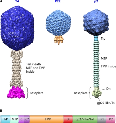
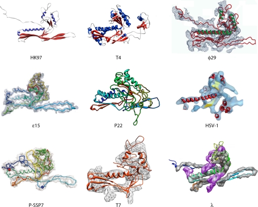
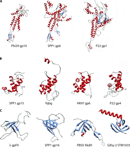
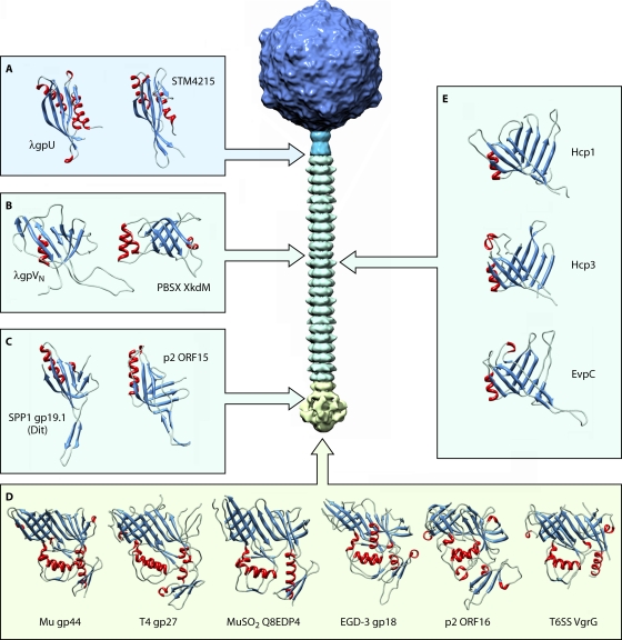
Bacteriophages belonging to the order Caudovirales possess a tail acting as a molecular nanomachine used during infection to recognize the host cell wall, attach to it, pierce it, and ensure the high-efficiency delivery of the genomic DNA to the host cytoplasm. In this review, we provide a comprehensive analysis of the various proteins constituting tailed bacteriophages from a structural viewpoint. To this end, we had in mind to pinpoint the resemblances within and between functional modules such as capsid/tail connectors, the tails themselves, or the tail distal host recognition devices, termed baseplates. This comparison has been extended to bacterial machineries embedded in the cell wall, for which shared molecular homology with phages has been recently revealed. This is the case for the type VI secretion system (T6SS), an inverted phage tail at the bacterial surface, or bacteriocins. Gathering all these data, we propose that a unique ancestral protein fold may have given rise to a large number of bacteriophage modules as well as to some related bacterial machinery components.


Figures

Fig. 1.
Fig. 1.
(A) The three Caudovirales families. From left to right are the Myoviridae (T4), the Podoviridae (P22), and the Siphoviridae (p2). (B) Schematic representation of the typical genome organization within the Siphoviridae tail morphogenesis module (this organization is also observed for several myophages with some adaptations). Trp, tail terminator; MTP, major tail protein; C and C*, tail chaperones; TMP, tape measure protein; Dit, distal tail protein; gp27-like/Tal (tail-associated lysozyme or tail fiber), the presence of a C-terminal domain depends on the phage considered; P1 and P2, baseplate/tip peripheral proteins (their number varies among phages).
Fig. 2.
Fig. 2.
The HK97 lineage. Shown is a gallery of HK97-like capsid protein structures determined by using crystallography for HK97 gp5 and T4 gp24 or cryo-EM for ϕ29 gp5, ε15 gp7, P22 gp5, herpes simplex virus type 1 (HSV-1) VP5, P-SSP7 gp10, T7 gp10, and λ gpE. (The HK97, T4, and HSV-1 images were adapted from reference by permission from Macmillan Publishers Ltd.; the ϕ29 image was adapted from reference with permission of the publisher; the ε15 image was adapted from reference with permission from Macmillan Publishers Ltd.; the P22 image was adapted from reference with permission of the publisher; the P-SSP7 image was adapted from reference with permission of Macmillan Publishers Ltd.; the T7 image was adapted from reference with permission of the publisher; and the λ image was adapted from reference with permission of the publisher.)
Fig. 3.
Fig. 3.
Conservation of the protein modules constituting bacteriophage connectors. (A) Gallery of portal protein structures determined by X-ray diffraction: ϕ29 gp10 (PDB accession no. 1FOU), SPP1 gp6 (PDB accession no. 2JES), and P22 gp1 (PDB accession no. 3LJ4). (B) Crystal structures of SPP1 gp15 (PDB accession no. 2KBZ), PBSX YqbG (PDB accession no. 1XN8), HK97 gp6 (PDB accession no. 3JVO), and P22 gp4 (PDB accession no. 3LJ4). (C) Crystal structures of λ gpFII (PDB accession no. 1K0H), SPP1 gp16 (PDB accession no. 2KCA), PBSX XkdH (PDB accession no. 3F3B), and Gifsy-2 STM1035 (PDB accession no. 2PP6). The coloring scheme used is based on secondary structures: blue, β-strands; red, α-helices; cyan, loops.
Fig. 4.
Fig. 4.
Conservation of the protein modules constituting bacteriophage tails and the T6SS apparatus needle. (A) Crystal structures of the tail terminator proteins λ gpU (PDB accession no. 3FZ2) and PBSX XkdM (PDB accession no. 2GJV). (B) Structures of the tail tube proteins of siphophage λ (gpVN [PDB accession no. 2K4Q]) and myophage PBSX (XkdM [PDB accession no. 2GUJ]) (C) Crystal structures of the Dit proteins SPP1 gp19.1 (PDB accession no. 2X8K) and p2 ORF15 (PDB accession no. 2WZP). (D) Gallery of gp27-like protein structures observed for the Myoviridae, including Mu gp44 (PDB accession no. 1WRU), T4 gp27 (PDB accession no. 1K28), and MuSO2 Q8EDP4 (PDB accession no. 3CDD); for the Siphoviridae, including EGD-e gp18 (PDB accession no. 3GS9) and p2 ORF16 (PDB accession no. 2WZP); as well as for the E. coli T6SS, including CFT073 VgrG (PDB accession no. 2P5Z). (E) Phage tail tube-like proteins from the T6SS tube: Hcp1 (PDB accession no. 1Y12) and Hcp3 (PDB accession no. 3HE1) from Pseudomonas aeruginosa and EVPC from Edwardsiella tarda (PDB accession no. 3EAA).

References

    1. Agirrezabala X., et al. 2007. Quasi-atomic model of bacteriophage t7 procapsid shell: insights into the structure and evolution of a basic fold. Structure 15:461–472 - PubMed
    1. Aksyuk A. A., et al. 2009. The tail sheath structure of bacteriophage T4: a molecular machine for infecting bacteria. EMBO J. 28:821–829 - PMC - PubMed
    1. Auzat I., Droge A., Weise F., Lurz R., Tavares P. 2008. Origin and function of the two major tail proteins of bacteriophage SPP1. Mol. Microbiol. 70:557–569 - PubMed
    1. Baker M. L., Jiang W., Rixon F. J., Chiu W. 2005. Common ancestry of herpesviruses and tailed DNA bacteriophages. J. Virol. 79:14967–14970 - PMC - PubMed
    1. Ballister E. R., Lai A. H., Zuckermann R. N., Cheng Y., Mougous J. D. 2008. In vitro self-assembly of tailorable nanotubes from a simple protein building block. Proc. Natl. Acad. Sci. U. S. A. 105:3733–3738 - PMC - PubMed

Publication types