Review
. 2011 Sep;75(3):434-67, first page of table of contents.
doi: 10.1128/MMBR.00008-11.

Molecular mechanism of scanning and start codon selection in eukaryotes

Affiliations
Review

Molecular mechanism of scanning and start codon selection in eukaryotes

Alan G Hinnebusch. Microbiol Mol Biol Rev. 2011 Sep.

Abstract

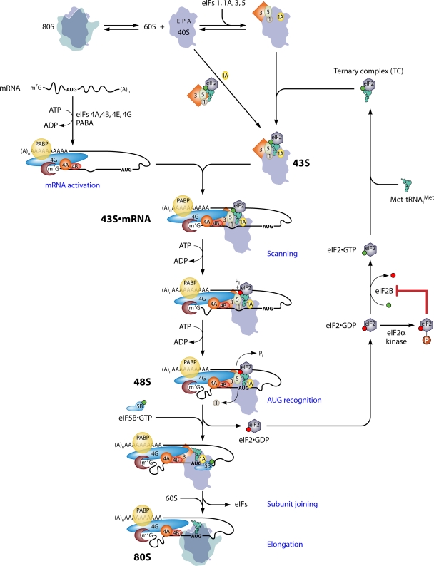
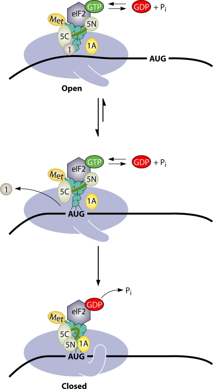
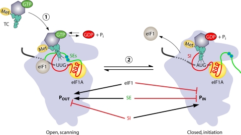
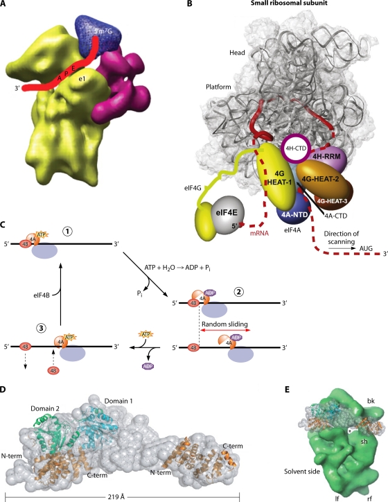
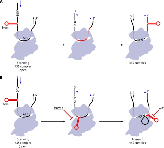
The correct translation of mRNA depends critically on the ability to initiate at the right AUG codon. For most mRNAs in eukaryotic cells, this is accomplished by the scanning mechanism, wherein the small (40S) ribosomal subunit attaches to the 5' end of the mRNA and then inspects the leader base by base for an AUG in a suitable context, using complementarity with the anticodon of methionyl initiator tRNA (Met-tRNAiMet) as the key means of identifying AUG. Over the past decade, a combination of yeast genetics, biochemical analysis in reconstituted systems, and structural biology has enabled great progress in deciphering the mechanism of ribosomal scanning. A robust molecular model now exists, describing the roles of initiation factors, notably eukaryotic initiation factor 1 (eIF1) and eIF1A, in stabilizing an "open" conformation of the 40S subunit with Met-tRNAiMet bound in a low-affinity state conducive to scanning and in triggering rearrangement into a "closed" conformation incompatible with scanning, which features Met-tRNAiMet more tightly bound to the "P" site and base paired with AUG. It has also emerged that multiple DEAD-box RNA helicases participate in producing a single-stranded "landing pad" for the 40S subunit and in removing the secondary structure to enable the mRNA to traverse the 40S mRNA-binding channel in the single-stranded form for base-by-base inspection in the P site.


Figures

Fig. 1.
Fig. 1.
Pathway of eukaryotic translation initiation via ribosomal scanning. The process of initiation is depicted as a pathway of reactions (a subset described in blue type), beginning with the dissociation of 80S ribosomes into free 40S and 60S subunits and the assembly of the 43S PIC on the small ribosomal subunit. 80S ribosomes and 40S subunits are depicted as silhouettes of the crystal structures of bacterial 70S and 30S ribosomal species, with approximate locations of the aminoacyl-tRNA (A), peptidyl-tRNA (P), and exit (E) sites labeled in the 40S subunit. eIFs are depicted as shapes labeled by numbers, and GTP or GDP bound to eIF2 in the ternary complex (TC) is depicted as green or red balls, respectively. Two pathways for the recruitment of the TC to the 40S subunit in assembling the 43S PIC are depicted, with one involving the prior incorporation of the TC into the multifactor complex (MFC). mRNA is activated by the binding of eIF4F (eIF4E/eIF4G/eIF4A) to the m7G cap and of PABP to the poly(A) tail of the mRNA, with the attendant production of a single-stranded region at the mRNA 5′ end in a manner facilitated by eIF4B and the ATP-dependent helicase activity of eIF4A. 43S PICs attach to the 5′ end of the mRNA, forming the 43S·mRNA complex, in a manner stabilized by a network of interactions among the mRNA, eIF4G, eIF3, eIF5, eIF4B, and the 40S subunit. The 43S PIC scans the mRNA 5′UTR in a manner facilitated by eIF4A's helicase activity, and the helicases Ded1 and Dhx29 (not shown) also facilitate scanning through stable secondary structures in the 5′UTR. Note that the hydrolysis of the GTP in the TC occurs in the scanning complex, but the release of Pi is blocked until the AUG base pairs with the anticodon of Met-tRNAiMet, with the attendant dissociation of eIF1 from the 40S subunit and the arrest of scanning. eIF2-GDP is released from Met-tRNAiMet, and eIF5B-GTP joins the complex to catalyze the joining of the 60S subunit and the production of an 80S initiation complex competent for elongation. The eIF2-GDP is recycled to the eIF2-GTP by the GEF eIF2B to enable the reassembly of the TC. eIF2B is inhibited by the phosphorylation of eIF2 on Ser-51 of its α subunit by various kinases, activated by different kinds of stress. See the text for more details. (Modified from reference with permission of Cold Spring Harbor Laboratory Press.)
Fig. 2.
Fig. 2.
Path of mRNA on the bacterial 70S ribosome. (A) Details of the arrangement of mRNA (yellow/orange/red/blue winding cylinder) relative to the major landmarks of the 30S subunit. The mRNA enters and exits on the solvent face of the 30S subunit (not shown here) and winds around the neck, being exposed on the interface surface of the subunit at the A, P, and E decoding sites. (Modified slightly from reference with permission from Elsevier.) (B) Toeprinting analysis of the bacterial PIC containing Met-tRNAiMet base paired to AUG in the P site. The view is looking down from above the head. Primer extension by reverse transcriptase (RT) from a 32P-labeled DNA primer (blue) annealed to the mRNA (black) downstream of the PIC is inhibited when RT collides with the 30S subunit near the entry channel opening, producing a cDNA (red) ∼17 nt smaller than the distance between the 5′ end of the primer and the AUG. (Adapted from reference with kind permission from Springer Science+Business Media.)
Fig. 3.
Fig. 3.
Schematic model of GCN4 translational control and mechanisms of Gcd and Gcn mutations. (A) Following the translation of uORF1 (boxed 1), posttermination 40S subunits remain attached to the GCN4 mRNA and resume scanning. Under nonstarvation conditions (left), they quickly rebind the TC and reinitiate at uORF4 (boxed 4), and the 80S ribosome dissociates after terminating at uORF4. Under amino acid starvation conditions (right), the concentration of the TC is reduced by eIF2α phosphorylation, such that many 40S ribosomes fail to rebind the TC until scanning past uORF4 and can reinitiate at the GCN4 ORF instead. (B) GCN4 translation is normally constitutively repressed in gcn2Δ cells owing to the inability to phosphorylate eIF2α in response to starvation. However, mutations that reduce the rate of TC loading on 40S subunits, such as substitutions in the eIF1A SE elements (eIF1A-SE*) (see text for details), constitutively derepress GCN4 translation, producing the Gcd phenotype. (C, left) Defects in different steps of reinitiation pictured here all prevent the induction of GCN4 translation in starved cells despite the reduction in TC levels, conferring the Gcn phenotype. Mechanisms of Gcn phenotypes can be discerned by analyzing the expression of the solitary uORF1 constructs depicted on the right. Gcn mutations that confer slow scanning should increase reinitiation at GCN4 for construct I, in which uORF1 is very close to the GCN4 uORF, by increasing the scanning time available for the reassembly of the PIC, but have little effect on construct II, in which uORF1 is far upstream of GCN4, and the scanning time available for reinitiation is long. Gcn mutations that confer the release of posttermination 40S subunits or reduce their ability to resume scanning will reduce the expression of constructs I and II. Gcn mutations that confer a leaky scanning of uORF1 will increase expression from construct III, in which uORF1 is elongated and extensively overlaps the GCN4 ORF, abolishing reinitiation.
Fig. 4.
Fig. 4.
Scheme for identifying Sui and Ssu mutations in yeast. See the text for details.
Fig. 5.
Fig. 5.
Cryo-EM model of the yeast eIF1·eIF1A·40S PIC. (A) Cryo-EM reconstructions of free 40S and the indicated complexes with eIF1 or eIF1A, which display moderately closed (apo-40S), open (40S-eIF1-eIF1A), or strongly closed (40S-eIF1A) conformations of the mRNA entry channel latch. (Reprinted from reference with permission from Elsevier.) (B) The images in A have been annotated here with schematics of the mRNA, eIF1A, eIF1, and the TC to summarize the biochemical findings (159) that the rate of TC binding is stimulated by both eIF1 and eIF1A but that the TC is bound more tightly to the PIC in the absence of eIF1, which would prevail with the eIF1 dissociation upon AUG recognition.
Fig. 6.
Fig. 6.
Schematic model depicting the structural rearrangements of the PIC thought to occur in the transition from the open conformation to the closed conformation upon AUG recognition. (Top) The presence of eIF1 and eIF1A on the 40S subunit stabilizes the open, scanning-conducive conformation of the PIC with an open conformation of the mRNA entry channel latch. TC binding is relatively unstable but in a configuration that allows the base-by-base inspection of the 5′UTR by Met-tRNAiMet in the P site. The GTP in the TC of a fraction of scanning complexes is hydrolyzed to eIF2·GDP·Pi, catalyzed by the N-terminal (GAP) domain of eIF5 (5N), but Pi release is blocked by eIF1. eIF1 maintains its association with the eIF5 CTD (5C), an interaction involved in stabilizing the yeast MFC. (Middle) AUG-Ac pairing evokes eIF1 dissociation from the 40S platform, and this enables isomerization to the closed conformation (Bottom), with the mRNA entry channel latch clamped on the mRNA. The absence of eIF1 in this state also enables tighter binding of TC to the P site and the completion of GTP hydrolysis and dissocation of Pi from eIF2·GDP·Pi. It was proposed that the absence of eIF1 allows the eIF5 NTD to switch partners from the G domain of eIF2γ to a 40S location that might overlap with the eIF1-binding site, enabling the eIF5 NTD interaction with eIF1A in a manner that stabilizes the closed conformation. eIF3 and the eIF4 group of factors were omitted from this diagram for simplicity. (Reproduced from reference with permission from Elsevier.)
Fig. 7.
Fig. 7.
Structure and functional domains of yeast eIF1. (Left) The amino acid sequences of the unstructured NTTs of yeast (y) and human (h) eIF1s are aligned and highlighted in green. The substitutions in −M1, −M2, and −M3 mutants of yeast eIF1 are shown below in red, which impair the interaction of eIF1 with the eIF2β NTD and eIF5 CTD in vitro. (Right) Ribbon model of yeast eIF1 indicating the positions of the −M4, −M5, and 93-97 (32) as well as the location of the “KH” and “KR” areas. The effects of the −M5 mutation on the interaction of eIF1 with the eIF3c NTD in vitro or native 40S subunits in vivo and of the −M4 mutation on eIF1 binding to the eIF2β NTD and the eIF5 CTD in vitro (180) are summarized. The interaction of α1 and the β1-β2 loop with the 40S subunit is predicted from structural studies (127, 178). (Reproduced from reference with permission of the publisher.)
Fig. 8.
Fig. 8.
Model depicting the opposite functions of the unstructured tails of eIF1A in modulating the conformational rearrangement of the PIC and distinct modes of TC binding to the open and closed states. The SE elements in the eIF1A CTT (shown in green) stabilize the open conformation of the PIC and the “Pout” mode of TC binding, which is compatible with scanning at non-AUG codons. The SEs also block the full P-site accommodation of initiator tRNA in the “Pin” mode required for start codon selection. eIF1 performs the same functions as the SE elements. The SI elements in the NTT and helical domain of eIF1A (both shown in red) function oppositely and destabilize the open conformation and the Pout mode of TC binding, thus promoting the closed conformation and Pin mode of initiator tRNA binding at AUG codons. Both the dissociation of eIF1 from the 40S subunit and the eviction of the SEs from the P site stabilize the closed conformation and tighter TC binding afforded by the Pin state.
Fig. 9.
Fig. 9.
Model of the 48S PIC containing bound eIF1, eIF1A, mRNA, and initiator tRNA. The model is based on directed hydroxyl radical mapping of mammalian eIF1 (127) and eIF1A (228) on 40S subunits and the crystal structure of bacterial 70S elongation complexes containing mRNA and P-site tRNA. The orientation of the tRNA and its location deep in the P site shown here might differ significantly from the eukaryotic scanning 43S or 48S PICs. The image in B is rotated 90° clockwise about the vertical axis from that in A, and the ribosomal protein S13/RpS18e in the head is not shown because it blocks the view of the eIF1A CTT. Ribbon representations of eIF1 are shown in lilac, and the eIF1A domains are shown in blue (OB fold), red (helical domain), green (NTT), and yellow (CTT). mRNA (gold) and tRNA (copper) are shown in the bottom panel only. (Reproduced from reference by permission of Oxford University Press.)
Fig. 10.
Fig. 10.
Substitution of 18S rRNA residues predicted to contact the P-site tRNA or P-site codon in elongating 80S ribosomes confers a phenotype indicating impaired TC binding to the scanning 43S PIC. Shown is the disposition of the bulge G926 of h28 (lilac in panels A and B) and various other 16S rRNA residues that contact the P-site codon or tRNA in a crystal structure of a bacterial 70S complex with mRNA and P-site tRNA. Substitutions of the corresponding residues in yeast 18S rRNA are lethal or confer an Slg phenotype and in most cases also evoke a dominant Gcd phenotype (detected in cells also expressing wild-type 18S rRNA), indicating less stable TC binding to the scanning PIC. (Modified from reference with permission of Cold Spring Harbor Laboratory Press.)
Fig. 11.
Fig. 11.
Interfaces between subunits of archaeal aIF2 and docking tRNA on aIF2γ. (A) Model of Phe-tRNAPhe (in a stick figure) docked onto the S. solfataricus aIF2αγ heterodimer. aIF2γ residues corresponding to those in yeast eIF2γ implicated in tRNA binding by the Sui substitutions Y142H and G397A are circled. See the text for more details. (Reproduced from reference with slight modifications, with permission from Elsevier.) (B and C) Segments of aIF2α, aIF2γ, and aIF2β at the subunit interfaces of the S. solfataricus heterotrimer of full-length β and γ subunits and domain III of aIF2α. Residues are labeled with a prefix (a, b, or g) indicating their subunit origin (α, β, or γ). (B) Helix α1 in aIF2β is wedged between two helices in the G domain of aIF2γ. The aIF2β residue corresponding to the yeast Sui substitution Y131 is circled. (C) sw1 in the G domain interacts directly with residues in the ZBD of aIF2β and with domain II of aIF2γ. The ZBD also contacts the ribose ring of the bound GDP (shown in a stick figure). Several ZBD residues near the interface with sw1 are immediately adjacent to residues corresponding to yeast eIF2β residues altered by Sui substitutions that confer increased GTP hydrolysis by the TC. (Panels B and C are reproduced from reference with slight modifications, with permission of the publisher [copyright 2007 National Academy of Sciences, U.S.A.].)
Fig. 12.
Fig. 12.
Models of the scanning PIC. (A) Cryo-EM model of mammalian eIF3 (magenta) and eIF4G (blue) binding to the 40S subunit (yellow), annotated to depict the path of the mRNA (red ribbon) in the decoding center and exit channel (Reproduced from reference with permission from AAAS.) (B) Model for the scanning PIC in which the eIF4G N-terminal region remains bound to the cap-eIF4E assembly and the HEAT-1 domain of eIF4G interacts with mRNA sequences immediately 5′ of the 40S exit channel, causing the mRNA to form a loop between the cap and HEAT-1 that grows in size as scanning proceeds. eIF4A, bound to HEAT-1, interacts with mRNA sequences located 3′ of the PIC to destabilize the secondary structure before it reaches the 40S subunit, and eIF4H prevents the reannealing of the unwound, single-stranded nucleotides before they enter the entry channel pore. (Reproduced from reference with permission from Elsevier.) (C) A Brownian ratchet mechanism of scanning in which eIF4A undergoes cycles of binding and dissociation from the mRNA, driven by ATP binding and hydrolysis, near the mRNA exit channel (where it is positioned by eIF4G). eIF4B interacts with the eIF4A·ATP·mRNA complex but dissociates from the mRNA more slowly than does eIF4A·ADP, allowing it to act as a pawl to prevent 3′-to-5′ backsliding. Thus, the random sliding of the ribosome can occur only in the 3′ direction and is fixed at the new location by the next cycle of eIF4A·ATP·mRNA·eIF4B complex assembly. (Reproduced from reference with permission of the publisher [copyright 2009 American Chemical Society].) (D, left) Docking of the crystal structure of yeast eIF4A to eIF4G and the HEAT-1 domain of human eIF4G1 into the SAXS envelope of the complex between eIF4A and the eIF4G fragment containing HEAT domains 1 to 3. (Right) Comparison of the sizes of the 40S subunit and the eIF4A-eIF4G complex. (Reproduced from reference by permission of Oxford University Press.)
Fig. 13.
Fig. 13.
Model depicting the bypass of a highly stable stem-loop by the scanning PIC in the presence of eIF4F but in the absence of helicase Dhx29. (A) A stem of moderate stability is unwound by eIF4F, eIF4A, and eIF4B and fed into the entry channel in a single-stranded form, producing a functional 48S PIC at the AUG. (B) A highly stable stem is not unwound effectively by the eIF4 factors alone, as this requires Dhx29 helicase function, bypasses the entry channel, and gets stuck in the exit channel. The mRNA spools into the decoding center, and a 48S PIC can be formed when AUG enters the P site provided that eIF1 is absent, as eIF1 destabilizes such aberrant complexes. (Reproduced from reference by permission from Macmillan Publishers Ltd., copyright 2011.)

References

    1. Abaeva I. S., Marintchev A., Pisareva V. P., Hellen C. U., Pestova T. V. 2011. Bypassing of stems versus linear base-by-base inspection of mammalian mRNAs during ribosomal scanning. EMBO J. 30:115–129 - PMC - PubMed
    1. Abastado J. P., Miller P. F., Jackson B. M., Hinnebusch A. G. 1991. Suppression of ribosomal reinitiation at upstream open reading frames in amino acid-starved cells forms the basis for GCN4 translational control. Mol. Cell. Biol. 11:486–496 - PMC - PubMed
    1. Acker M. G., Shin B. S., Dever T. E., Lorsch J. R. 2006. Interaction between eukaryotic initiation factors 1A and 5B is required for efficient ribosomal subunit joining. J. Biol. Chem. 281:8469–8475 - PubMed
    1. Agris P. F. 2008. Bringing order to translation: the contributions of tRNA anticodon-domain modifications. EMBO Rep. 9:629–635 - PMC - PubMed
    1. Algire M. A., Maag D., Lorsch J. R. 2005. Pi release from eIF2, not GTP hydrolysis, is the step controlled by start-site selection during eukaryotic translation initiation. Mol. Cell 20:251–262 - PubMed

MeSH terms

LinkOut - more resources