Review
. 2011 Sep;75(3):507-42, second and third pages of table of contents.
doi: 10.1128/MMBR.00009-11.

The universally conserved prokaryotic GTPases

Affiliations
Review

The universally conserved prokaryotic GTPases

Natalie Verstraeten et al. Microbiol Mol Biol Rev. 2011 Sep.

Abstract

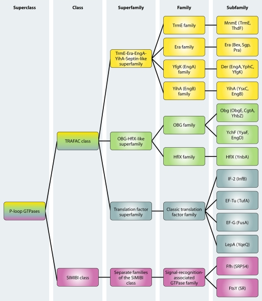
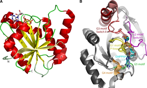
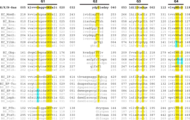
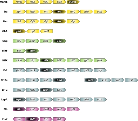
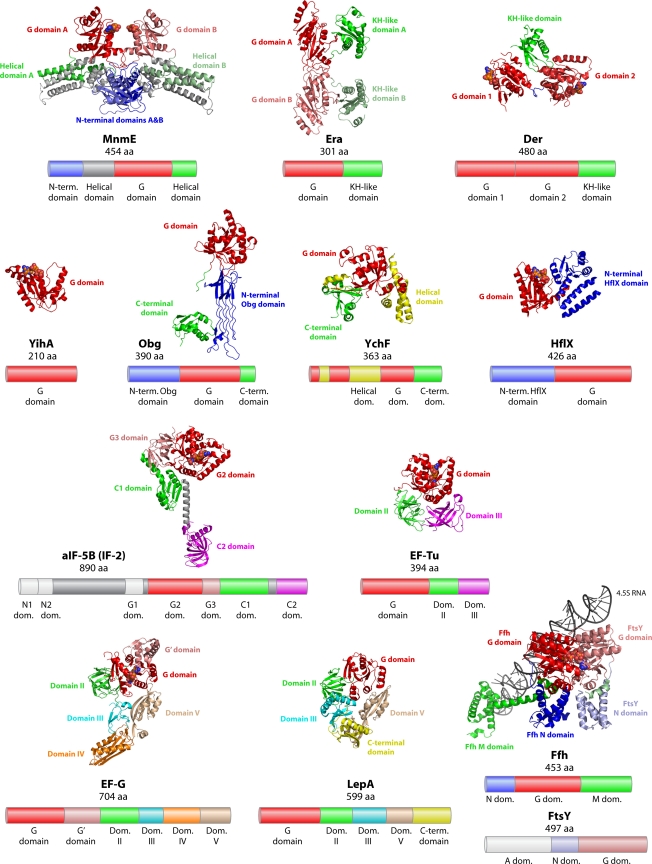
Members of the large superclass of P-loop GTPases share a core domain with a conserved three-dimensional structure. In eukaryotes, these proteins are implicated in various crucial cellular processes, including translation, membrane trafficking, cell cycle progression, and membrane signaling. As targets of mutation and toxins, GTPases are involved in the pathogenesis of cancer and infectious diseases. In prokaryotes also, it is hard to overestimate the importance of GTPases in cell physiology. Numerous papers have shed new light on the role of bacterial GTPases in cell cycle regulation, ribosome assembly, the stress response, and other cellular processes. Moreover, bacterial GTPases have been identified as high-potential drug targets. A key paper published over 2 decades ago stated that, "It may never again be possible to capture [GTPases] in a family portrait" (H. R. Bourne, D. A. Sanders, and F. McCormick, Nature 348:125-132, 1990) and indeed, the last 20 years have seen a tremendous increase in publications on the subject. Sequence analysis identified 13 bacterial GTPases that are conserved in at least 75% of all bacterial species. We here provide an overview of these 13 protein subfamilies, covering their cellular functions as well as cellular localization and expression levels, three-dimensional structures, biochemical properties, and gene organization. Conserved roles in eukaryotic homologs will be discussed as well. A comprehensive overview summarizing current knowledge on prokaryotic GTPases will aid in further elucidating the function of these important proteins.


Figures

Fig. 1.
Fig. 1.
Classification of GTPases described in this review. The superclass of P-loop GTPases is subdivided into two classes. The TRAFAC class is comprised of universally conserved protein families belonging to the TrmE-Era-EngA-YihA-septin-like superfamily (yellow), the OBG-HflX-like superfamily (green), and the translation factor superfamily (blue). From the SIMIBI class, only GTPases belonging to the signal-recognition-associated GTPase family (violet) are universally conserved. (Data adapted from reference .)
Fig. 2.
Fig. 2.
Overall structure of the G domain of P-loop GTPases. (A) Ribbon plot of a G domain. The structure of B. subtilis YihA in complex with GDP is shown (Protein Data Bank [PDB] accession number 1SVI). β-Strands are shown in yellow, α-helices are in red, and connecting loops are in green. GDP is shown in a stick representation. YihA contains an extra N-terminal β-strand and α-helix compared to the minimal 6-stranded mixed β-sheet of Ras. Conforming to the secondary structure numbering of Ras, these extra elements have been numbered β-strand and α-helix −1. A peptide region connecting α-helix 1 and β-strand 2 (corresponding to switch I) is disordered in this structure and is not shown. (B) Conserved GTPase motifs. The figure shows a superposition of B. subtilis YihA in the GDP-bound “off” state (PDB accession number 1SVI) and B. subtilis YihA bound to the GTP analog GMPPNP, mimicking the “on” state (PDB accession number 1SVW). The conserved sequence elements and the switch regions are shown in different colors, as indicated. YihA-GMPPNP is shown in dark-shaded colors, while YihA-GDP is shown in the corresponding lighter-shaded colors. In YihA-GDP, switch I and switch II are disordered and ordered, respectively, while in YihA-GMPPNP, switch I and switch II are ordered and disordered, respectively (216).
Fig. 3.
Fig. 3.
Sequence alignment of conserved G motifs. The amino acid sequence of human Ras G motifs is shown in boldface type. Other proteins are grouped according to their classifications in superfamilies (153). Sequences for E. coli (EC) and B. subtilis (BS) homologs are shown in black and gray, respectively. Residues conserved in all GTPases are highlighted in yellow. Residues deviating from the consensus sequence are marked in blue. The length of the G motifs was chosen as described previously (24). Sequences of the respective G2 motifs were obtained from previous work (24, 34, 43, 97, 115, 188, 207, 293).
Fig. 4.
Fig. 4.
Gene organization of E. coli GTPases described in this review. Color coding corresponds to the GTPase classification shown in Fig. 1. Genes encoding GTPases are indicated in light lettering on a dark background. The figure was constructed by using the Search Tool for the Retrieval of Interacting Genes (STRING) database (247). For YihA, no neighboring genes were identified by using STRING, and the gene organization is shown as described in reference .
Fig. 5.
Fig. 5.
Structures of GTPases described in this review. Structural domains are indicated and colored as follows: red, G domains; blue and gray, domains N terminal to the G domain; yellow, domains integrated into the G domain; green, magenta, violet, cyan, orange, brown, and yellow, domains C terminal to the G domain. Nucleotides bound to the active site of the GTPase are shown in a sphere representation. One representative of each subfamily is shown: MnmE, structure of MnmE from Nostoc sp. in complex with GDP (PDB accession number 3GEH); Era, structure of Era from E. coli in the apo form (PDB accession number 1EGA); Der, structure of Der from B. subtilis in complex with GDP (PDB accession number 2HJG); YihA, structure of YihA from B. subtilis in complex with GDP (PDB accession number 1SVI); Obg, Obg from T. thermophilus in the apo form (PDB accession number 1UDX); YchF, YchF from H. influenzae in the apo form (PDB accession number 1JAL); HflX, homolog of HflX from the archaeon S. solfataricus in complex with GDP (PDB accession number 2QTH); IF-2, homolog of IF-2 from the archaeon M. thermautotrophicus (aIF5b) in complex with GMPPNP (PDB accession number 1G7T); EF-Tu, EF-Tu from T. thermophilus in complex with GMPPNP (PDB accession number 1EXM); EF-G, slow mutant of EF-G from T. thermophilus in complex with GTP (PDB accession number 2BV3); LepA, LepA from E. coli in the apo form (PDB accession number 3CB4); Ffh-FtsY, E. coli Ffh in complex with E. coli FtsY and the 4.5S RNA from D. radiodurans, with both proteins bound to the nonhydrolyzable GTP analog β, γ-methylene-GTP (GMPPCP) (PDB accession number 2XXA). Below each structure, the domain structure of the corresponding E. coli homolog is shown with the corresponding color coding. Additional E. coli domains are marked in light gray. Pfam accession numbers are P25522 (MnmE), P06616 (Era), P0A6P5 (Der), P0A6P7 (YihA), P42641 (Obg), P0ABU2 (YchF), P25519 (HflX), P0A6N1 (EF-Tu), P0A6M8 (EF-G), P0A705 (IF-2), P60785 (LepA), P0AGD7 (Ffh), and P10121 (FtsY). aa, amino acids.
Fig. 6.
Fig. 6.
Uridine modification by MnmE. MnmA (in collaboration with other proteins) carries out thiolation at the 2-position of the wobble uridine, whereas an α2β2 heterotetrameric complex formed by MnmE and MnmG independently catalyzes the first step of the cmnm5 modification at the 5-position. MnmC has two enzymatic activities that transform the cmnm5 intermediate into the final mnm5 modification in certain bacteria. (Adapted from reference by permission of Oxford University Press.)
None
None
None
None

References

    1. Agirrezabala X., Frank J. 2009. Elongation in translation as a dynamic interaction among the ribosome, tRNA, and elongation factors EF-G and EF-Tu. Q. Rev. Biophys. 42:159–200 - PMC - PubMed
    1. Agris P. F. 2004. Decoding the genome: a modified view. Nucleic Acids Res. 32:223–238 - PMC - PubMed
    1. Ahnn J., March P. E., Takiff H. E., Inouye M. 1986. A GTP-binding protein of Escherichia coli has homology to yeast RAS proteins. Proc. Natl. Acad. Sci. U. S. A. 83:8849–8853 - PMC - PubMed
    1. Akiyama T., et al. 2001. Mammalian homologue of E. coli Ras-like GTPase (ERA) is a possible apoptosis regulator with RNA binding activity. Genes Cells 6:987–1001 - PubMed
    1. Alam K. Y., Clark D. P. 1991. Molecular cloning and sequence of the thdF gene, which is involved in thiophene and furan oxidation by Escherichia coli. J. Bacteriol. 173:6018–6024 - PMC - PubMed

Publication types

MeSH terms

Substances

LinkOut - more resources