. 2011 Sep 2;333(6047):1292-6.
doi: 10.1126/science.1206606.

Chemical and genetic engineering of selective ion channel-ligand interactions

Affiliations

Chemical and genetic engineering of selective ion channel-ligand interactions

Christopher J Magnus et al. Science. .

Abstract

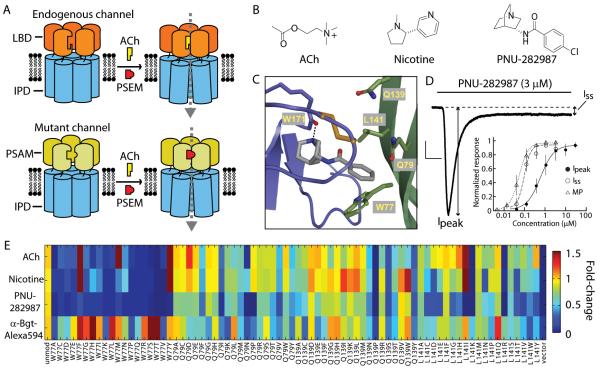
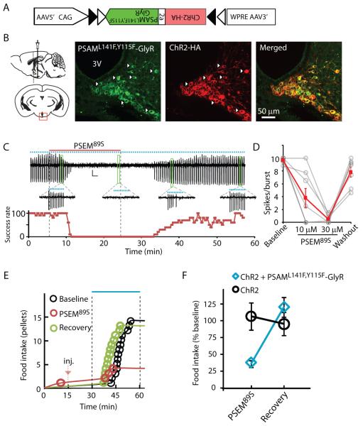
Ionic flux mediates essential physiological and behavioral functions in defined cell populations. Cell type-specific activators of diverse ionic conductances are needed for probing these effects. We combined chemistry and protein engineering to enable the systematic creation of a toolbox of ligand-gated ion channels (LGICs) with orthogonal pharmacologic selectivity and divergent functional properties. The LGICs and their small-molecule effectors were able to activate a range of ionic conductances in genetically specified cell types. LGICs constructed for neuronal perturbation could be used to selectively manipulate neuron activity in mammalian brains in vivo. The diversity of ion channel tools accessible from this approach will be useful for examining the relationship between neuronal activity and animal behavior, as well as for cell biological and physiological applications requiring chemical control of ion conductance.


Figures

Fig. 1
Fig. 1
“Bump-hole” approach to engineer selective ion channel-ligand interactions. (A) LGICs composed of ligand binding domain (LBD) and ion pore domain (IPD) modules. LBD mutations yield a Pharmacologically Selective Actuator Module (PSAM) that selectively binds Pharmacologically Selective Effector Molecules (PSEMs, red) but not the endogenous ligand (acetylcholine, ACh, yellow). PSEMs do not bind the unmodified LBD. (B) Chemical structures of ACh, nicotine, and PNU-282987. (C) Homology model of the α7 nAChR LBD with a docked agonist at the interface between two protomers (purple and green). Residues W77, Q79, Q139, and L141 were targeted for mutagenesis. (D) Current evoked by PNU-282987 application to an HEK 293 cell expressing α7-5HT3. A large peak current (Ipeak) rapidly decays to a persistent, steady state current (Iss). Black line indicates time course of ligand application. Scale bars: 200 pA, 0.5 s. Inset, Dose response curves (normalized to maximum response) from MP assay correspond to Iss (n = 3). (E) Color map showing activity and cell surface expression of mutated α7-5HT3 channels. Responses to ACh (100 μM), nicotine (100 μM), PNU-282987 (10 μM), and binding of α-Bungarotoxin (Bgt)-Alexa594 were normalized to the response of the unmodified channel to ACh (100 μM) or α-Bgt-Alexa594 binding. Error bars are s.e.m.
Fig. 2
Fig. 2
Selective interactions between ligands and mutated α7-5HT3 chimeric ion channels. (A) Color map showing EC50s for mutated α7-5HT3 with ACh, nicotine, and aminoquinuclidine benzamides. In order to highlight molecules that are highly selective for mutated α7-5HT3 chimeric receptors, only the activity of molecules with EC50 > 30 μM against unmodified α7-5HT3 (top row) are shown for the mutated receptors. (B) Dose response curves for compounds against mutated channels and showing negligible activation of unmodified α7-5HT3. EC50MP in parentheses. Normalization is to the maximum response to each compound or to ACh (for unmodified α7-5HT3). (C) Dose response curves for mutated ion channel-ligand interactions that show orthogonal pharmacology. Normalization is to maximum response for each compound across mutated channels. (D) Chemical structures of ligands in B and C. Error bars are s.e.m.
Fig. 3
Fig. 3
PSAM/IPD chimeric ion channels for neuron activation and silencing. (A) Confocal projection image of α-Bgt-Alexa594 labeling in a cortical brain slice expressing PSAMQ79G,Q139G-5HT3 HC receptors. Left, Laminar boundaries. (B) Depolarization of layer 2/3 cortical neurons (in tetrodotoxin and CNQX) via PSAM-5HT3 HC channels by PSEMs applied at 10 μM. PSEM application (30 μM) to control neurons electroporated with GFP-expressing vector (green) did not depolarize cells. Sample sizes in parentheses. (C) Cell attached recordings of PSAM-5HT3 HC channel activation of layer 2/3 cortical neurons. Scale bar: 50 pA. Ligand application, 120 s. (D) Ligand selectivity: layer 2/3 cortical neurons expressing PSAML141F,Y115F-5HT3 HC are activated by PSEM89S but not PSEM22S. (E) Top, Confocal projection image of α-Bgt labeling in a cortical brain slice expressing PSAML141F,Y115F-GlyR receptors. Bottom, dendritic segment. (F,G) PSAML141F,Y115F-GlyR and PSEM89S (10 μM, black, n = 16), but not PSEM89S alone (30 μM, white, n = 10), reversibly reduce cellular input resistance, Rin. Shown in (G) is Rin normalized to pre-PSEM89S application (PRE); values are displayed for PSEM89S application and after 3 min wash-out (WASH). (H) PSAML141F,Y115F-GlyR reversibly reduces excitability of cortical neurons to depolarizing current injection in the presence of PSEM89S. (I) Rheobase, normalized to pre-PSEM89S application. Error bars are s.e.m.
Fig. 4
Fig. 4
Stringent neuronal silencing test of PSAML141F,Y115F-GlyR for suppressing AGRP neuron-evoked feeding behavior. (A) Construct for a Cre-dependent recombinant adeno-associated viral vector with an inverted bicistronic open reading frame for PSAML141F,Y115F-GlyR and ChR2 under control of a FLEX-switch [two antiparallel pairs of heterotypic loxP sites (triangles)]. (B) Diagram illustrating injection site (left) and confocal images (right) taken from brain slices of virally transduced Agrp-cre mice showing co-expression of PSAML141F,Y115F-GlyR (green, α-Bgt-Alexa488) and ChR2 (red, anti-HA). (C) Cell-attached recording showing suppression of light-evoked bursts of action potential currents (10 Hz light pulses for 1 s, represented by blue dots) following application and then wash-out of PSEM89S (above). Bursts are expanded for selected time points. Below is spike success rate (percent) before, during and after PSEM89S application. (D) Successful light-evoked spikes per burst (n = 8 cells). (E) Food intake on successive days resulting from photostimulation in an Agrp-cre mouse co-expressing ChR2 and PSAML141F,Y115F-GlyR after saline (Baseline and Recovery) or PSEM89S [intraperitoneal injection (inj.), marked with red arrow]. (F) Evoked food intake normalized to initial photostimulation session. Injection of PSEM89S reduced photostimulation-evoked eating in Agrp-cre mice co-expressing ChR2 and PSAML141F,Y115F-GlyR (30 mg/kg, blue diamond, n = 5 mice) but not in Agrp-cre mice expressing only ChR2 (50 mg/kg, black circle, n = 6 mice). Error bars are s.e.m.

Comment in

References

    1. Slimko EM, McKinney S, Anderson DJ, Davidson N, Lester HA. Selective electrical silencing of mammalian neurons in vitro by the use of invertebrate ligand-gated chloride channels. J. Neurosci. 2002;22:7373. - PMC - PubMed
    1. Tan EM, et al. Selective and quickly reversible inactivation of mammalian neurons in vivo using the Drosophila allatostatin receptor. Neuron. 2006;51:157. - PubMed
    1. Wulff P, et al. From synapse to behavior: rapid modulation of defined neuronal types with engineered GABAA receptors. Nat. Neurosci. 2007;10:923. - PMC - PubMed
    1. Armbruster BN, Li X, Pausch MH, Herlitze S, Roth BL. Evolving the lock to fit the key to create a family of G protein-coupled receptors potently activated by an inert ligand. Proc. Natl. Acad. Sci. U. S. A. 2007;104:5163. - PMC - PubMed
    1. Gosgnach S, et al. V1 spinal neurons regulate the speed of vertebrate locomotor outputs. Nature. 2006;440:215. - PubMed

Publication types

MeSH terms