. 2011 Nov;60(11):2701-9.
doi: 10.2337/db11-0489. Epub 2011 Sep 1.

CCK stimulation of GLP-1 neurons involves α1-adrenoceptor-mediated increase in glutamatergic synaptic inputs

Affiliations

CCK stimulation of GLP-1 neurons involves α1-adrenoceptor-mediated increase in glutamatergic synaptic inputs

Kazunari Hisadome et al. Diabetes. 2011 Nov.

Abstract

Objective: Glucagon-like peptide 1 (GLP-1) is involved in the central regulation of food intake. It is produced within the brain by preproglucagon (PPG) neurons, which are located primarily within the brain stem. These neurons project widely throughout the brain, including to the appetite centers in the hypothalamus, and are believed to convey signals related to satiety. Previous work demonstrated that they are directly activated by leptin and electrical activity of the afferent vagus. Another satiety hormone, cholecystokinin (CCK), has also been linked to activation of brain stem neurons, suggesting that it might act partially via centrally projecting neurons from the nucleus tractus solitarius (NTS). The aim of this study was to investigate the neuronal circuitry linking CCK to the population of NTS-PPG neurons.

Research design and methods: Transgenic mice expressing yellow fluorescent protein (Venus) under the control of the PPG promoter were used to identify PPG neurons in vitro and to record their electrical and pharmacological profile.

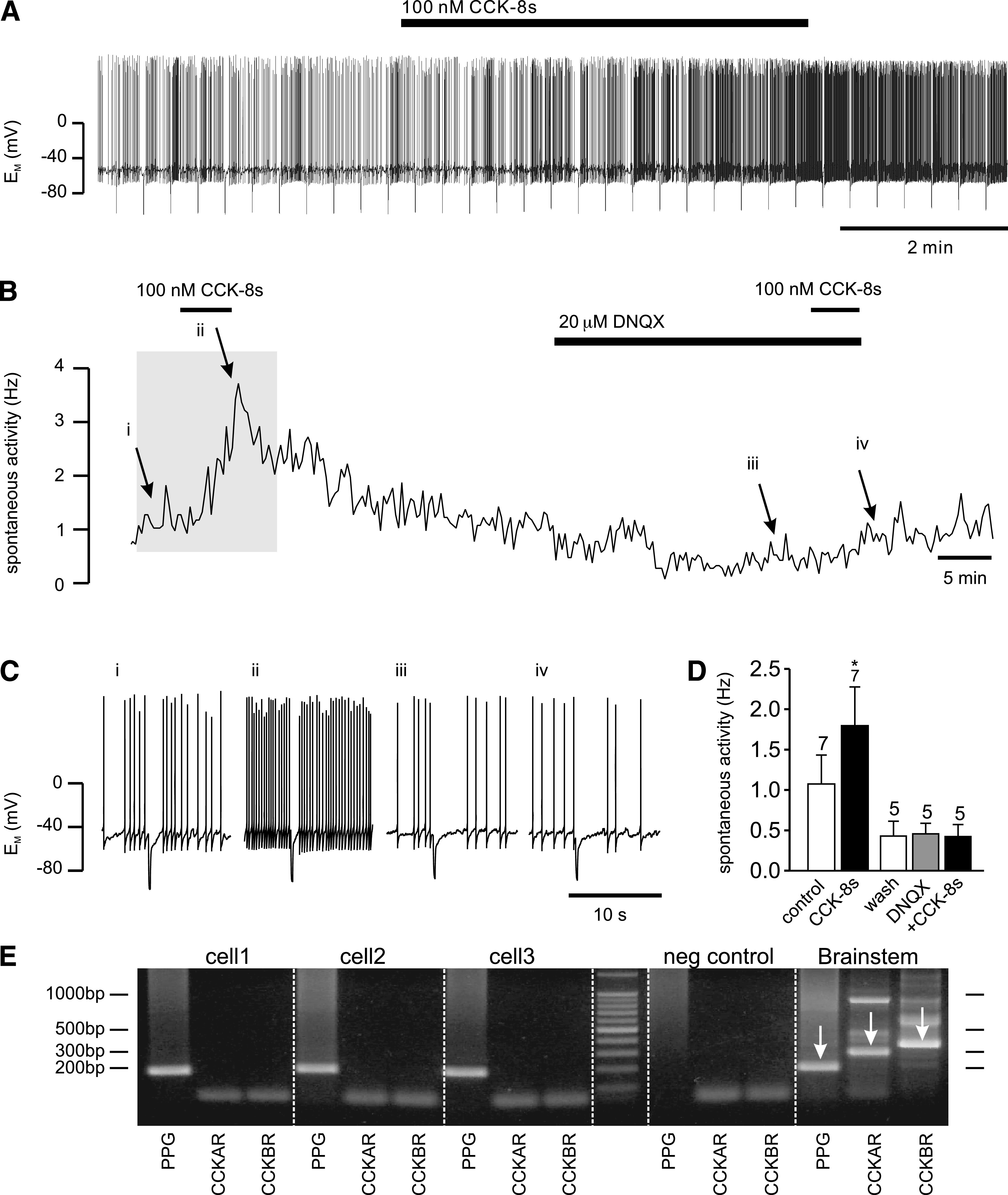
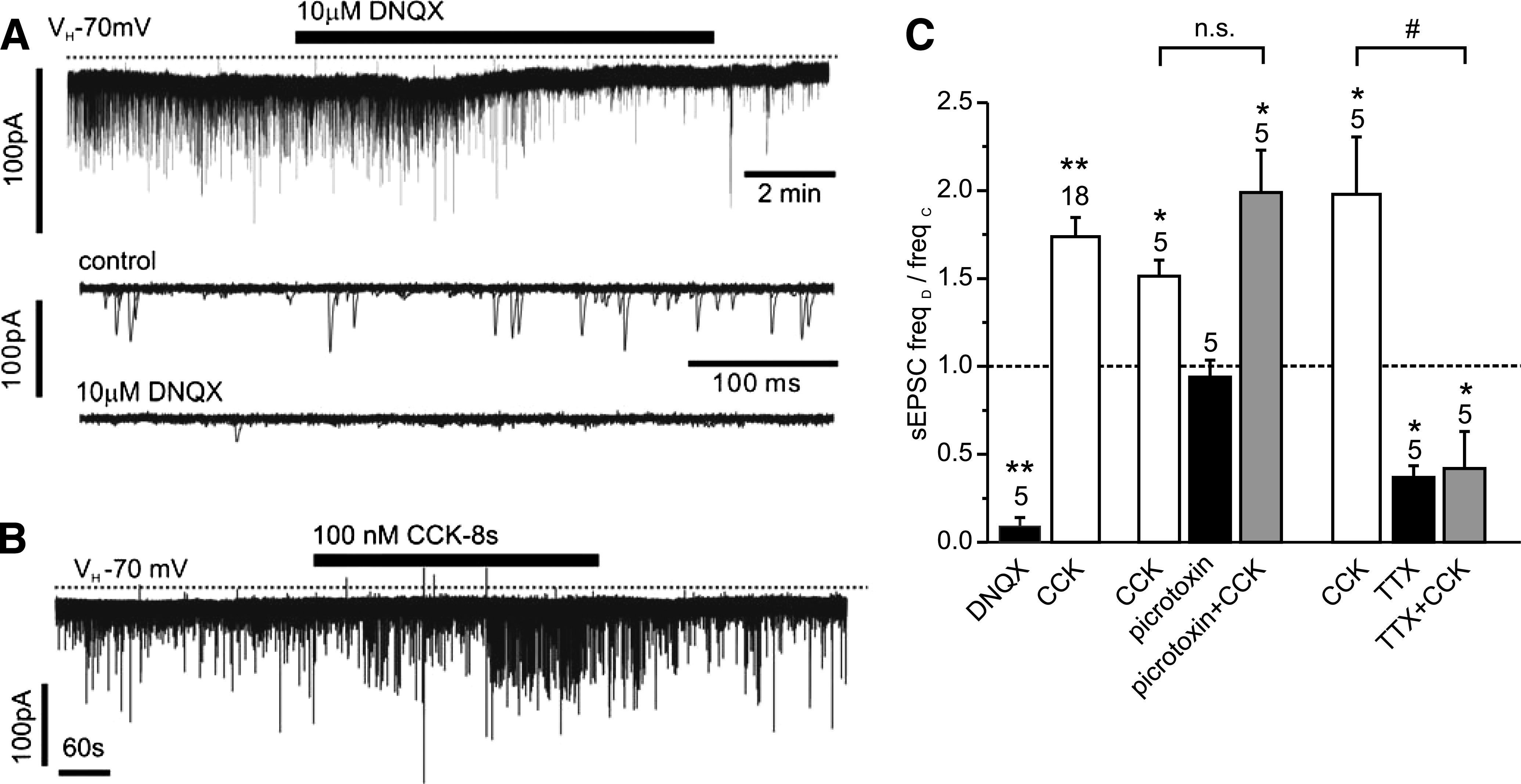
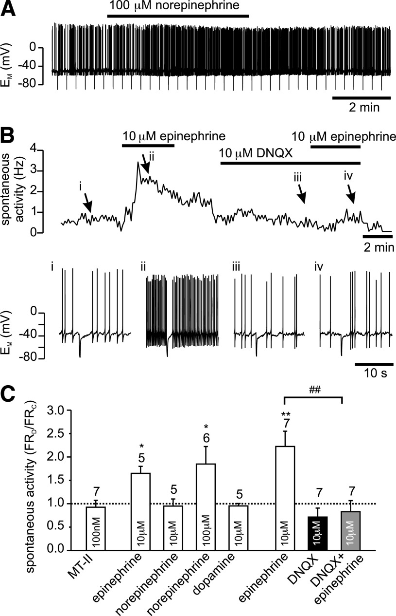
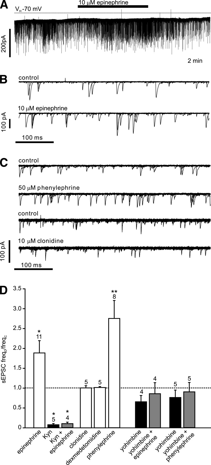
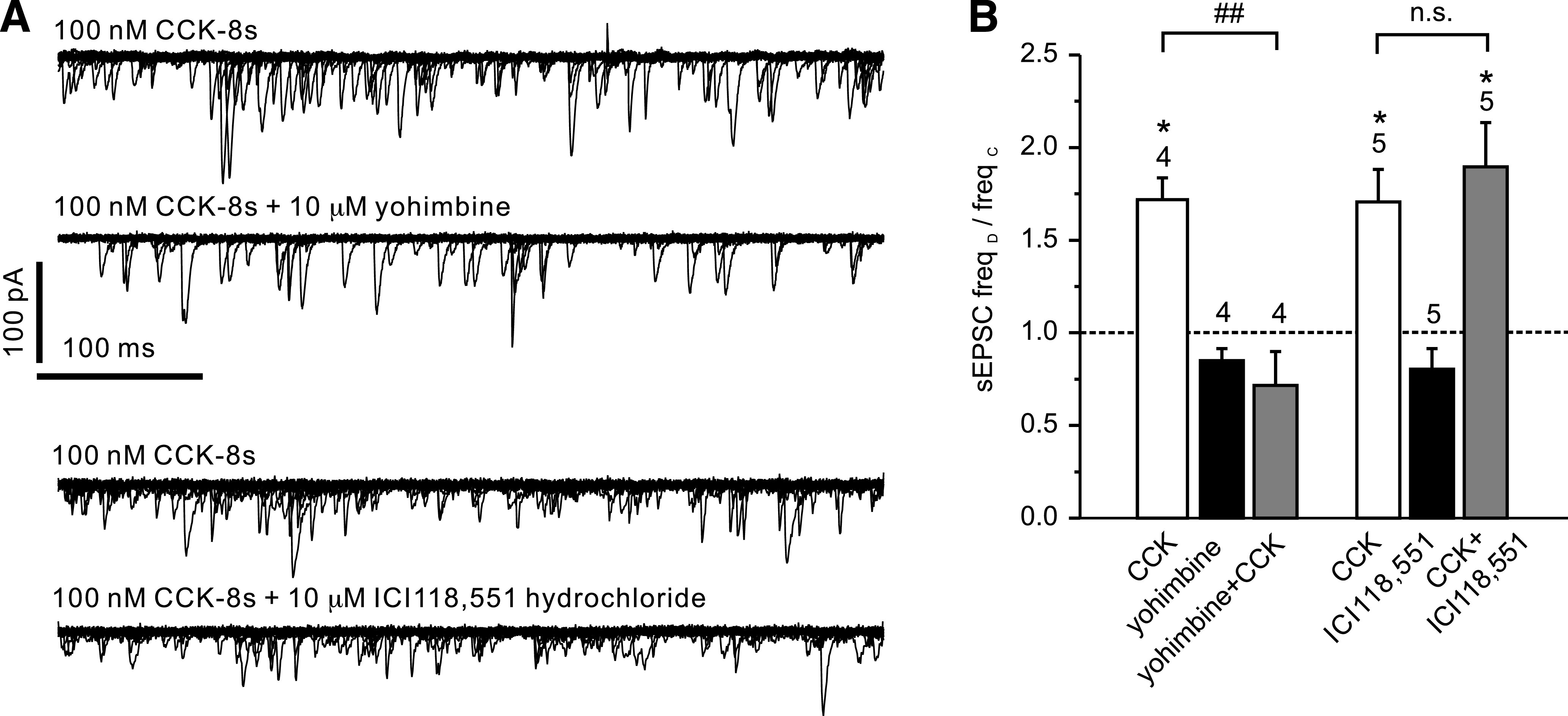
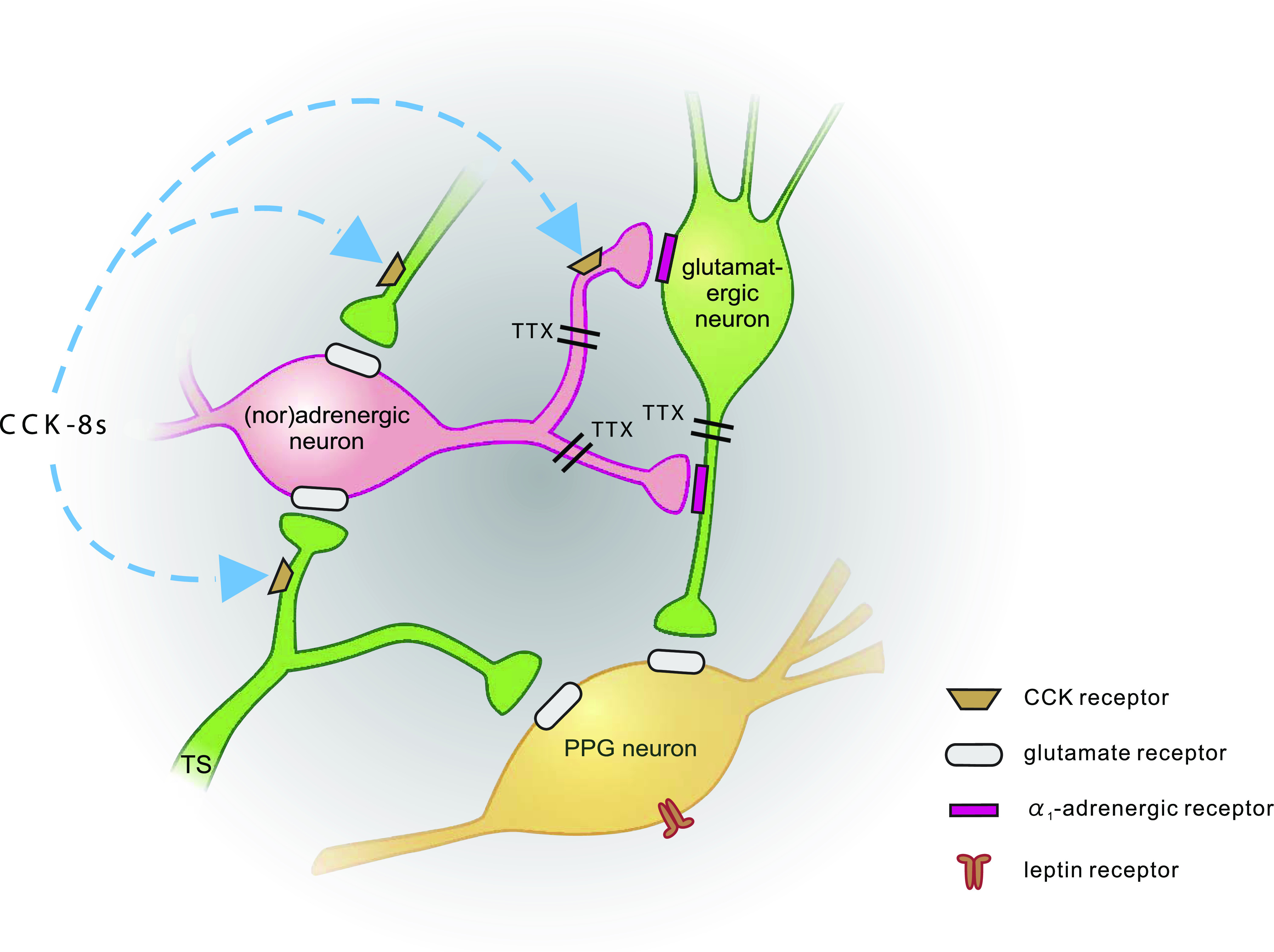
Results: PPG neurons in the NTS were excited by CCK and epinephrine, but not by the melanocortin receptor agonist melanotan II. Both CCK and epinephrine acted to increase glutamatergic transmission to the PPG neurons, and this involved activation of α(1)-adrenergic receptors. Inhibition of adrenergic signaling abolished the excitatory action of CCK.

Conclusions: CCK activates NTS-PPG cells by a circuit involving adrenergic and glutamatergic neurons. NTS-PPG neurons integrate a variety of peripheral signals that indicate both long-term energy balance and short-term nutritional and digestional status to produce an output signal to feeding and autonomic circuits.


Figures

FIG. 1.
FIG. 1.
CCK increases spontaneous activity of PPG neurons. A: Current-clamp recording demonstrating that bath application of 100 nmol/L CCK-8s led to an increase in spontaneous action potential firing frequency of this PPG neuron. B: A plot of the firing frequency for the recording shown in A (the part of the recording shown in A is indicated by gray background). C: Short segments of the original current-clamp recording from B at time points indicated by i, ii, iii, and iv. D: Mean data for firing frequency from experiments as depicted in A and B. CCK-8 (100 nmol/L) significantly increased firing rate. This effect of CCK-8s was occluded in the presence of the non-NMDA glutamate receptor antagonist DNQX. Number of recordings for each condition is given above the bars. *P < 0.05. E: Typical single-cell RT-PCR analysis for PPG and the CCK receptors (CCKAR, CCKBR) for three PPG neurons and controls. Agarose gel (2%) demonstrating that the 186-bp PCR product for PPG, the 285-bp PCR product for CCKAR, and the 341-bp product for CCKBR can be obtained from brain stem cDNA (1:100 dilution; positive control; indicated by arrows) with the primers specified in Table 1. In contrast, cytoplasm extracted from single cells showing eYFP fluorescence (cell1, cell2, cell3) was only positive for PPG, but not CCKAR or CCKBR (only bands for primers visible). Negative (neg) control: pipette solution without cytoplasm extracted from cell. Molecular weight ladder shows bands at 100-bp intervals.
FIG. 2.
FIG. 2.
CCK stimulation of sEPSCs is sensitive to TTX but not picrotoxin. A: The vast majority of sEPSCs in PPG neurons are glutamatergic, as demonstrated by their inhibition by 10 μmol/L DNQX in this voltage-clamp recording at a holding potential of −70 mV. Bottom traces: Overlay of 15 consecutive 500 ms traces from the recording above under control conditions (top) and in the presence of DNQX (bottom). B: CCK-8s, bath-applied at 100 nmol/L, led to an increase in sEPSC frequency. C: Mean normalized effects of CCK on sEPSC frequency in the presence or absence of various drugs. The mean sEPSC frequency in presence of the drug (freqD) as a fraction of the frequency in the absence of any drug (freqC) is plotted. The excitatory CCK effect is not reduced by the GABA and glycine receptor antagonist picrotoxin (30 μmol/L) but is prevented by TTX (0.5 μM). *P < 0.05 compared with control; **P < 0.01 compared with control; #P < 0.05 compared with CCK. Numbers of cells tested are given above the bars.
FIG. 3.
FIG. 3.
Epinephrine stimulation of firing frequency of PPG neurons is occluded by DNQX. A: Current-clamp recording showing the effect of bath application of 100 μmol/L norepinephrine on firing frequency of PPG neuron. B: Bath application of 10 μmol/L epinephrine leads to an increase in spontaneous action potential firing frequency of PPG neurons. This effect of epinephrine is prevented by the non-NMDA glutamate receptor antagonist DNQX (10 μmol/L). Top: instantaneous firing frequency; bottom: segments of the original current-clamp recording at time points i, ii, iii, and iv, indicated by arrows. C: Mean data for the change in firing frequency (FR) during experiments as shown in A. Epinephrine (10 μmol/L) and 100 μmol/L norepinephrine, but not 100 nmol/L Melanotan II (MT-II), 10 μmol/L norepinephrine, or 10 μmol/L dopamine significantly increased firing rate. The effect of 10 μmol/L epinephrine is occluded by 10 μmol/L DNQX. Number of recordings for each condition is given above the bars. *P < 0.05, **P < 0.01, compared with control; ##P < 0.01 compared with epinephrine.
FIG. 4.
FIG. 4.
Epinephrine acts on α1-adrenoreceptors and increases the frequency of spontaneous glutamatergic EPSCs. A: Voltage-clamp recording from a PPG neuron at a holding potential (VH) of −70 mV demonstrating the effects of epinephrine on sEPSCs. B: Overlay of 15 consecutive 500 ms traces from the recording shown in A under control conditions (top) and in the presence of epinephrine (bottom). C: Overlay of 15 consecutive 500 ms traces under control conditions and in the presence of phenylephrine or clonidine, respectively, as indicated above each overlay. Phenylephrine, but not clonidine, led to an increase in sEPSC frequency. D: Mean normalized effects of epinephrine and selective α1- (phenylephrine) and α2- (clonidine, dexmedetomidine) adrenoreceptor agonists on sEPSC frequency. The mean sEPSC frequency in presence of the drug (freqD) as a fraction of the frequency in the absence of any drug (freqC) is plotted. The effect of epinephrine is blocked by the glutamate receptor antagonist kynurenic acid (Kyn) and by the α-adrenergic receptor antagonist yohimbine. Yohimbine also blocked the effect of phenylephrine. Kyn itself blocked the majority of sEPSCs. *P < 0.05, **P < 0.01, compared with control. Numbers of cells tested are given above the bars.
FIG. 5.
FIG. 5.
CCK stimulation of spontaneous EPSCs is sensitive to yohimbine. A: Overlay of 15 consecutive 500 ms traces from a recording like that shown in Fig. 2B in the presence of CCK (top) and CCK after preincubation with yohimbine (second from top). CCK-8s, bath-applied at 100 nmol/L, led to an increase in sEPSC frequency. This effect could be blocked by 10 μmol/L yohimbine. Bottom two traces show overlays from a recording where CCK was first applied alone (top trace) and then in the presence of the β-adrenoreceptor antagonist ICI118,551 hydrochloride (10 μmol/L; bottom trace). The CCK effect was not reduced by the β-adrenoreceptor antagonist. B: Mean normalized effects of CCK in the presence or absence of various drugs on sEPSC frequency from recordings as depicted in A. The mean sEPSC frequency in presence of the drug (freqD) as a fraction of the frequency in the absence of any drug (freqC) is plotted. The effect of CCK is blocked by yohimbine (10 μmol/L) but not ICI118,551. *P < 0.05 compared with control; ##P < 0.01 compared with CCK. Numbers of cells tested are given above the bars.
FIG. 6.
FIG. 6.
Schematic representation of synaptic inputs to PPG neurons. PPG neurons (yellow) express leptin receptors (Hisadome et al. [19]) and receive direct glutamatergic input both from the tractus solitarius (TS; TTX-insensitive; Hisadome et al. [(19)]) and local glutamatergic neurons (green; TTX-sensitive). Input from adrenergic/noradrenergic neurons (pink) is indirect via α1-adrenergic receptors, activation of which enhances glutamatergic input to PPG neurons. CCK enhances the activity of PPG neurons. Its effect is occluded by either α-adrenergic receptor antagonists or non-NMDA glutamate receptor antagonists. Thus, it acts either on (nor)adrenergic cells or presynaptic from those, as suggested by Baptista et al. (37).

References

    1. Holst JJ. The physiology of glucagon-like peptide 1. Physiol Rev 2007;87:1409–1439 - PubMed
    1. Trapp S, Hisadome K. Glucagon-like peptide 1 and the brain: central actions-central sources? Auton Neurosci 2011;161:14–19 - PubMed
    1. Merchenthaler I, Lane M, Shughrue P. Distribution of pre-pro-glucagon and glucagon-like peptide-1 receptor messenger RNAs in the rat central nervous system. J Comp Neurol 1999;403:261–280 - PubMed
    1. Jin SL, Han VK, Simmons JG, Towle AC, Lauder JM, Lund PK. Distribution of glucagonlike peptide I (GLP-I), glucagon, and glicentin in the rat brain: an immunocytochemical study. J Comp Neurol 1988;271:519–532 - PubMed
    1. Llewellyn-Smith IJ, Reimann F, Gribble FM, Trapp S. Preproglucagon neurons project widely to autonomic control areas in the mouse brain. Neuroscience 2011;180:111–121 - PMC - PubMed

Publication types

MeSH terms