. 2012 Mar;19(3):451-60.
doi: 10.1038/cdd.2011.113. Epub 2011 Sep 2.

A dp53/JNK-dependant feedback amplification loop is essential for the apoptotic response to stress in Drosophila

Affiliations

A dp53/JNK-dependant feedback amplification loop is essential for the apoptotic response to stress in Drosophila

E Shlevkov et al. Cell Death Differ. 2012 Mar.

Abstract

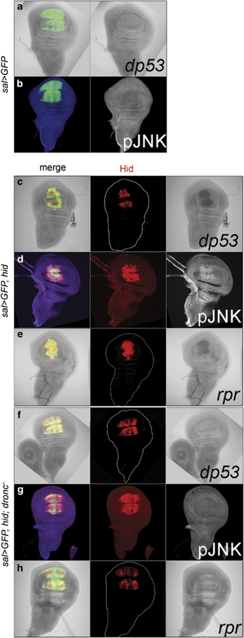
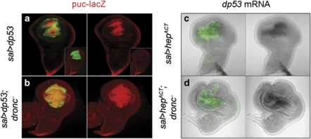
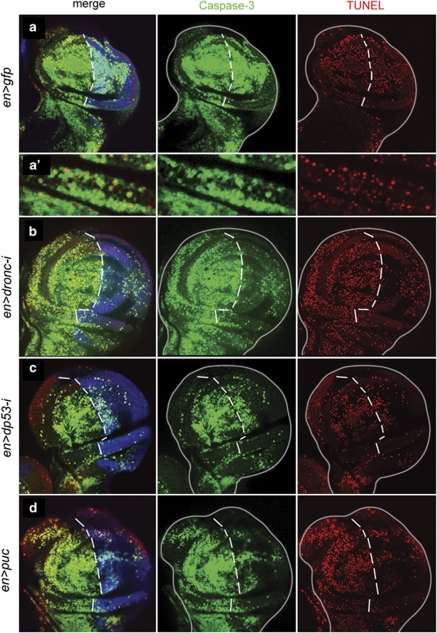
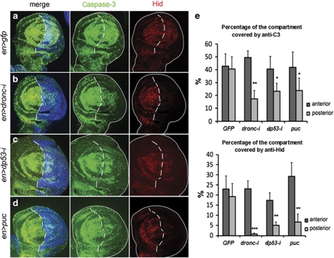
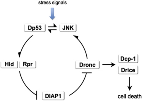
Programmed cell death (apoptosis) is a conserved process aimed to eliminate unwanted cells. The key molecules are a group of proteases called caspases that cleave vital proteins, which leads to the death of cells. In Drosophila, the apoptotic pathway is usually represented as a cascade of events in which an initial stimulus activates one or more of the proapoptotic genes (hid, rpr, grim), which in turn activate caspases. In stress-induced apoptosis, the dp53 (Drosophila p53) gene and the Jun N-terminal kinase (JNK) pathway function upstream in the activation of the proapoptotic genes. Here we demonstrate that dp53 and JNK also function downstream of proapoptotic genes and the initiator caspase Dronc (Drosophila NEDD2-like caspase) and that they establish a feedback loop that amplifies the initial apoptotic stimulus. This loop plays a critical role in the apoptotic response because in its absence there is a dramatic decrease in the amount of cell death after a pulse of the proapoptotic proteins Hid and Rpr. Thus, our results indicate that stress-induced apoptosis in Drosophila is dependant on an amplification loop mediated by dp53 and JNK. Furthermore, they also demonstrate a mechanism of mutual activation of proapoptotic genes.


Figures

Figure 1
Figure 1
Proapoptotic genes are activated by Dp53 and the JNK pathway. (a) Simplified scheme of the apoptotic pathway in Drosophila. Upstream activators like Dp53 and the JNK pathway induce one or more of the proapoptotic genes (only hid and rpr are shown in the scheme but there are other related genes, grim and sickle). The inactivation of the apoptosis inhibitor Diap1 by the proapoptotic proteins allows the proteolytic activation of the Dronc, Dcp-1 and Drice caspases and subsequent cell death (see Hay and Guo for a more detailed description). (b and c) Control discs of genotype spaltEPv-Gal4>UAS-GFP doubly stained for GFP and Hid and for GFP and rpr transcripts. There is neither hid nor rpr activity in the sal domain, labelled green. (dg) Activation of hid and rpr by Dp53 and JNK pathway. (d and e) Discs of genotype spaltEPv-Gal4>UAS-GFP UAS-dp53 showing Hid protein (d) and rpr transcription (e) in the sal domain. (f and g) Disc of genotype spaltEPv-Gal4>UAS-GFP UAS-hepACT showing accumulation of the Hid protein (f) and rpr activity (g) in the sal domain. (hk) The activation of hid and rpr by Dp53 and JNK pathway in dronc mutant discs. (h) Disc of genotype spaltEPv-Gal4>UAS-GFP UAS-dp53 dronc demonstrating the presence of the Hid protein in the expanded sal domain. (i) Disc of the same genotype as in (h) also showing rpr transcription in the expanded sal domain. (j and k) spaltEPv-Gal4>UAS-GFP UAS-hepACTdronc discs also showing gain of Hid (j) and rpr (k) in the sal domain. The sal domain in the dronc mutant discs becomes enlarged because of the ectopic expression of wg and dpp induced by JNK22
Figure 2
Figure 2
The proapoptotic factor Hid activates dp53, the JNK pathway and rpr in a dronc-dependant manner. (a and b) Control discs of genotype spaltEPv-Gal4>UAS-GFP doubly stained for GFP and dp53 transcript (a) and for GFP and the phosphorylated form of JNK/Basket (b). (c and d) The effect of Hid on dp53 transcription and JNK pathway activity. (c) spaltEPv-Gal4>UAS-GFP UAS-hid disc showing dp53 expression, revealed by in situ hybridization, in cells expressing hid, labelled by anti-Hid in red. (d) In the spaltEPv-Gal4>UAS-GFP UAS-hid disc, there is also JNK activity in the sal domain in coexpression with Hid, as revealed with the antibody that recognizes the phosphorylated form of JNK/Basket (gray). The (e) row illustrates the activation of rpr by Hid. Following the same scheme as in the first two rows, forcing expression of hid in the sal domain results in rpr activation, revealed by in situ hybridization. (f–h) The result of forcing hid in dronc discs is shown. (f) Disc of genotype spaltEPv-Gal4>UAS-GFP UAS-hid dronc showing Hid expression in the sal domain but no dp53 transcription. The (g) row demonstrates that JNK activity is absent in the spaltEPv-Gal4>UAS-GFP UAS-hid dronc disc, and (h) that in dronc mutant discs rpr remains silent
Figure 3
Figure 3
dp53 and JNK are able to activate each other independently of dronc function. (a and b) In sal>dp53 (spaltEPv-Gal4>UAS-dp53 UAS-GFP; puc-lacZ/+) and sal>dp53 dronc (spaltEPv-Gal4>UAS-dp53 UAS-GFP; pucE69-lacZ dronc124/dronc129) discs, the JNK pathway becomes activated, as revealed by the puc-LacZ marker. In otherwise wild-type discs, puc-lacZ expression is restricted to the most proximal cells (inset). Conversely, driving JNK expression in the sal domain activates dp53 transcription in both dronc+ and dronc discs (c and d)
Figure 4
Figure 4
Apoptotic response to a brief pulse of hid activity. (a) en-Gal4 >UAS-GFP; hs-hid wing disc showing high levels of anti-Caspase-3 (green) and TUNEL (red). GFP staining is shown in blue. A magnification is shown in (a′) to appreciate the concordance between anti-Caspase-3 and TUNEL staining. (b) en-Gal4>UAS-GFP UAS-dronc RNA-i; hs-hid showing a much reduced response in the posterior compartment. In the discs of genotype en-Gal4>UAS-GFP UAS-dp53 RNA-i; hs-hid (c) or en-Gal4>UAS-GFP UAS-puc; hs-hid (d), the apoptotic response in the posterior compartments is also much reduced. Note that TUNEL and anti-Caspase-3 staining are highly concordant in all cases
Figure 5
Figure 5
Loop-mediated amplification of Hid levels after a pulse of hid. (a) Control en-Gal4>UAS-GFP; hs-hid flies. The expression of GFP is shown in blue. The 30 min heat shock induces high levels of Caspase-3 (green) and Hid (red), both in the anterior and posterior compartments. (b) en-Gal4>UAS-GFP UAS-dronc RNA-i; hs-hid disc showing a large reduction of Caspase-3 and Hid activity in the posterior compartment because of the suppression of dronc function. The third panel clearly indicates that most of the Hid protein visible after the heat shock is generated by the feedback loop. (c) A en-Gal4> UAS-GFP UAS-dp53 RNA-i; hs-hid disc showing reduction of Caspase-3 and Hid in the posterior compartment, where dp53 function is diminished. (d) en-Gal4> UAS-GFP UAS-puc; hs-hid disc with reduced levels of Caspase-3 and Hid in the posterior compartment. (e) The results of quantitative measurements of Caspase-3 and Hid activities in the genotypes studied. The percentage of the area of each compartment covered by the staining with anti-Caspase-3 or anti-Hid was calculated as indicated in the Materials and Methods section. There is no statistically significant difference between the values in UAS-GFP-expressing discs (n=23, P>0.05 both for anti-Caspase-3 and anti-Hid). A statistically significant reduction in the values of the posterior compartment is observed when UAS-dronc-RNA-i (n=19, P<0.0001 for both markers), UAS-dp53-RNA-i (n=20, P<0.01 for anti-Caspase-3 and P<0.001 for anti-Hid) or UAS-puc (n=27, P<0.01 for anti-Caspase-3 and P<0.001 for anti-Hid) are crossed to en-Gal4 UAS-GFP flies. *P<0.01, **P<0.001, ***P<0.0001
Figure 6
Figure 6
Activation of the endogenous hid gene after a pulse of Hid protein. (aa″) Disc of the genotype en-Gal4>UAS-GFP, UAS-dronc RNA-i, hid05014-lacZ; hs-hid fixed 4 h after the end of the heat shock, labelled for β-gal (red) and caspase (green). The posterior compartment is labelled in blue. The panel (a') shows lacZ activity in the anterior compartment, which is largely coextensive with caspase activity, shown in (a″). (b) Non-heat shocked disc of genotype hs-hid/hid20−10-lacZ showing background levels of lacZ expression. (c and c′) Disc of the same genotype fixed 4 h after heat shock. lacZ expression is induced and is coextensive with high levels of caspase activity
Figure 7
Figure 7
Activation of the JNK pathway after physiological stress. (a) Control pucE69-lacZ/+ disc showing high levels of puc expression and caspase activity in the wing pouch 4 h after an irradiation of 2000R. The expression of puc in the proximal region (arrow) is normally present and corresponds to the midline cells. (b) Disc of genotype droncI24, pucE69-lacZ/droncI29 showing very low expression of puc and caspase activity after the same dose of irradiation as in (a). Note (arrow) the normal puc expression in the midline cells. (c) Disc of genotype en>diap1; pucE69-lacZ/+. The low dronc activity in the posterior compartment results in low level of puc expression and caspase activity after a 2000R radiation. (d) The left panel shows a quantification of puc levels (see Materials and Methods) in a normal nonirradiated pucE69-lacZ/+ disc, in an irradiated disc of the same phenotype and in an irradiated droncI24, pucE69-lacZ/droncI29 disc. The middle and right panels show amplification of the inset in the corresponding photographs in (c). **P<0.001
Figure 8
Figure 8
Amplification loop model of stress-induced apoptosis. We envisage stress-induced apoptosis as follows: an original stress event (irradiation, heat shock) induces dp53 function, which activates hid and rpr transcription and also the JNK pathway. The JNK pathway is also able to amplify dp53 transcription (and probably to induce rpr and hid as well). The activation of dronc because of Hid/Rpr function gives rise, on one hand, to the activation of effector caspases and, on the other, to an increase of both Dp53 and JNK levels, thus completing the amplification loop. This feedback is necessary for the completion of the apoptotic program, because in its absence there is a dramatic reduction in the amount of cell death. The model implies that the cell killing function of Dronc requires amplification of its own levels. This is achieved by its ability to induce dp53/JNK function. The model incorporates the functional interactions among the various factors involved, but it does not contemplate their interactions at the molecular level. Some of the molecular interactions between Hid, Rpr, Diap1, Dronc and Drice are well known,, but the mechanisms of activation of dp53 and JNK by Dronc and the specific molecular interactions between dp53 and JNK have not yet been described

References

    1. Jacobson MD, Weil M, Raff MC. Programmed cell death in animal development. Cell. 1997;88:347–354. - PubMed
    1. Meier P, Finch A, Evan G. Apoptosis in development. Nature. 2000;407:796–801. - PubMed
    1. Thornberry NA, Lazebnik Y. Caspases: enemies within. Science. 1998;281:1312–1316. - PubMed
    1. Reed JC, Doctor KS, Godzik A. The domains of apoptosis: a genomics perspective. Sci STKE. 2004;2004:re9. - PubMed
    1. Shiozaki EN, Shi Y. Caspases, IAPs and Smac/DIABLO: mechanisms from structural biology. Trends Biochem Sci. 2004;29:486–494. - PubMed

Publication types

MeSH terms

LinkOut - more resources