Review
. 2011 Sep 2;12(10):553-69.
doi: 10.1038/nrn3086.

Linking neural activity and molecular oscillations in the SCN

Affiliations
Review

Linking neural activity and molecular oscillations in the SCN

Christopher S Colwell. Nat Rev Neurosci. .

Abstract

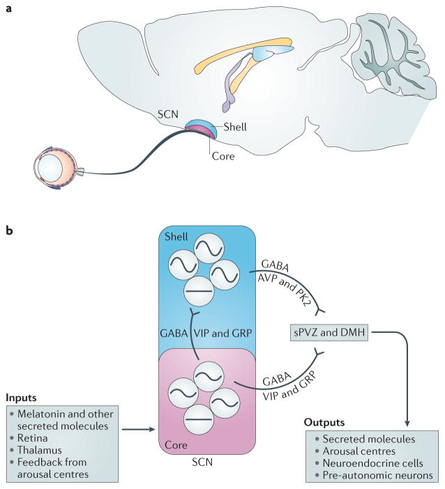
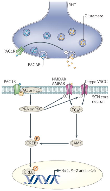
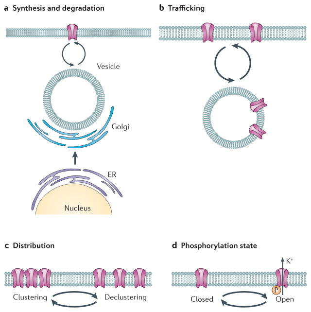
Neurons in the suprachiasmatic nucleus (SCN) function as part of a central timing circuit that drives daily changes in our behaviour and underlying physiology. A hallmark feature of SCN neuronal populations is that they are mostly electrically silent during the night, start to fire action potentials near dawn and then continue to generate action potentials with a slow and steady pace all day long. Sets of currents are responsible for this daily rhythm, with the strongest evidence for persistent Na(+) currents, L-type Ca(2+) currents, hyperpolarization-activated currents (I(H)), large-conductance Ca(2+) activated K(+) (BK) currents and fast delayed rectifier (FDR) K(+) currents. These rhythms in electrical activity are crucial for the function of the circadian timing system, including the expression of clock genes, and decline with ageing and disease. This article reviews our current understanding of the ionic and molecular mechanisms that drive the rhythmic firing patterns in the SCN.


Conflict of interest statement

Competing interests statement

The author declares no competing financial interests.

Figures

Figure 1
Figure 1. The suprachiasmatic nucleus circuit
a | The suprachiasmatic nucleus (SCN) in the hypothalamus is often divided into two anatomical and functional subdivisions: a ventrolateral ‘core’ and a dorsomedial ‘shell’. b | SCN core neurons are thought to integrate external input, including light information from the retinohypothalamic tract (RHT) and information from the thalamus and from midbrain structures such as the raphe nucleus. Core neurons relay this information to the rest of the SCN using GABA and vasoactive intestinal peptide (VIP) or gastrin-releasing peptide (GRP). Shell neurons use GABA and arginine vasopressin (AVP) or prokineticin 2 (PK2) to communicate with other cell populations, and at least some of the SCN shell neurons are neurosecretory cells that rhythmically release signalling molecules, including AVP, into the third ventricle. The amplitudes of the rhythms in gene expression and neural activity in core and shell neurons are relatively low and high, respectively. The outputs of core and shell SCN neurons travel mainly to other hypothalamic regions, including the subparaventricular zone (sPVZ) and the dorsal medial hypothalamus (DMH). These hypothalamic relay nuclei send projections throughout the CNS and endocrine system. Major centres in the brain that control arousal, such as the raphe nucleus, locus coeruleus, hypocretin or orexin neurons, and pars tuberalis, are rhythmically regulated through projections from the SCN.
Figure 2
Figure 2. How light regulates the molecular clockwork in SCN neurons
The best understood pathways by which neural activity regulates clock gene expression comes from studies that have explored how light turns on the transcription of period 1 (Per1) during the night. Melanopsin-expressing retinal ganglion cells encode ambient light and generate action potentials that travel down the retinohypothalamic tract (RHT) and innervate the suprachiasmatic nucleus (SCN). The RHT terminals release glutamate and, under certain conditions, the neuropeptide pituitary adenylate cyclase activating peptide (PACAP). The net result of RHT stimulation is an increase in firing rate and Ca2+ increase in SCN neurons through both activation of glutamate receptors (AMPA and/or NMDA receptors) and voltage-sensitive calcium currents (VSCCs). So far, all of the available evidence indicates that the PACAP type I receptor (PAC1R; also known as PACAPR1) is responsible for mediating the effects of PACAP on SCN neurons. Functionally, PACAP presynaptically enhances the release of glutamate onto SCN neurons and postsynaptically enhances the magnitude of NMDA and AMPA currents within the SCN. The increase in Ca2+ activates a number of signalling pathways that converge to alter transcriptional and/or translational regulators, including cyclic AMP-responsive element (CRE)-binding protein (CREB). Phosphorylated CREB is translocated into the nucleus where it can bind to CREs in the promoter regions of c-Fos, period 1 (Per1) and Per2, and drives transcription of these genes over the course of hours. Within the SCN, the photic regulation of c-Fos and Per1 is rapid, whereas the regulation of Per2 exhibits a slower time course. AC, adenylyl cyclase; CAMK, calcium/calmodulin-dependent protein kinase; PKA, protein kinase A; PKC, protein kinase C; PLC, phospholipase C.
Figure 3
Figure 3. Possible mechanisms by which the molecular clock can regulate spontaneous neural activity in SCN neurons
a | Ion channels and other membrane proteins are made and then transported to the membrane through the rough endoplasmic reticulum (ER) to the Golgi. The membrane proteins are transported to the membrane and removed for degradation through vesicles. There is evidence of the rhythmic transcription of several ion channels, including L- and T-type Ca2+ channels, large-conductance Ca2+ activated K+ (BK) channels and two-pore K+ (K2P) channels. Although we do not know the half-life of these channels in the SCN, direct transcription and translational regulation of membrane proteins is an obvious candidate mechanism for driving the observed rhythms in spontaneous neural activity. b | AMPA receptors and potassium channels have been shown to be rapidly inserted and removed from the membrane in response to physiological stimulation. Therefore, the daily trafficking of ion channels and associated proteins is another possible mechanism that may be responsible for the firing rate rhythms. c | The distribution of ion channels within the plasma membrane can change from day to night, and this may also contribute to changes in firing rate rhythms between day and night. d | Robust circadian rhythms in signalling pathways, including pathways involving Ca2+, cyclic AMP, phospholipase C (PLC), casein kinases and RAS-dependent mitrogen-activated protein kinases (MAPKs), have been described. These pathways are robust regulators (through phosphorylation) of ion channels and associated proteins. Circadian regulation in the phosphorylation state of ion channels through the balance between kinase and phosphotase activities is a likely mechanism that underlies the rhythm in spontaneous neural activity in SCN neurons. In summary, a diverse set of mechanisms, including changes in transcription and translation, direct phosphorylation, trafficking and distribution of ion channels (and associated proteins (not shown)) could underlie the rhythms in membrane events that characterize SCN neurons.

References

    1. Reppert SM, Weaver DR. Coordination of circadian timing in mammals. Nature. 2002;418:935–941. - PubMed
    1. Ko CH, Takahashi JS. Molecular components of the mammalian circadian clock. Hum Mol Genet. 2006;15:R271–R277. - PubMed
    1. Dibner C, Schibler U, Albrecht U. The mammalian circadian timing system: organization and coordination of central and peripheral clocks. Annu Rev Physiol. 2010;72:517–549. - PubMed
    1. Herzog ED. Neurons and networks in daily rhythms. Nature Rev Neurosci. 2007;8:790–802. - PubMed
    1. Welsh D, Takahashi J, Kay S. Suprachiasmatic nucleus: cell autonomy and network properties. Annu Rev Physiol. 2010;72:551–577. This review focuses on what is known about how cell-autonomous oscillators are coupled within the SCN circuit. - PMC - PubMed

Publication types