Review
. 2011 Sep 2;12(10):643-55.
doi: 10.1038/nrm3184.

Dynamic niches in the origination and differentiation of haematopoietic stem cells

Affiliations
Review

Dynamic niches in the origination and differentiation of haematopoietic stem cells

Leo D Wang et al. Nat Rev Mol Cell Biol. .

Erratum in

  • Nat Rev Mol Cell Biol. 2011;13(1):12

Abstract

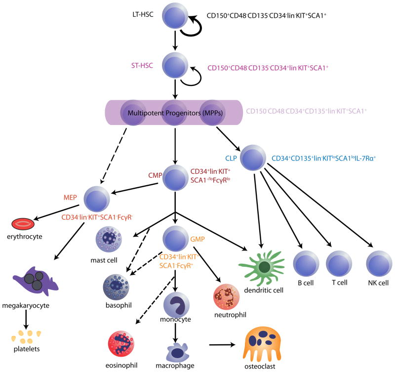
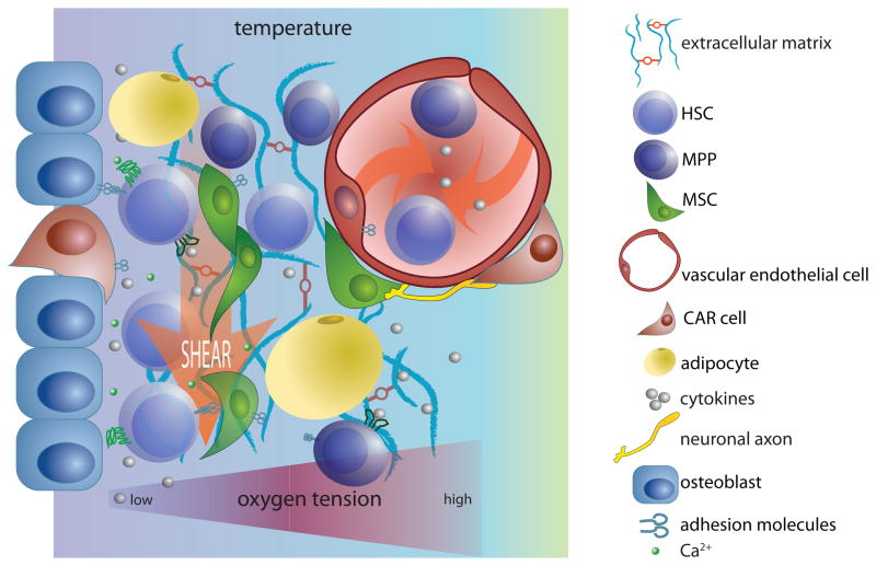
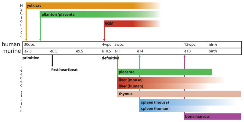
Haematopoietic stem cells (HSCs) are multipotent, self-renewing progenitors that generate all mature blood cells. HSC function is tightly controlled to maintain haematopoietic homeostasis, and this regulation relies on specialized cells and factors that constitute the haematopoietic 'niche', or microenvironment. Recent discoveries, aided in part by technological advances in in vivo imaging, have engendered a new appreciation for the dynamic nature of the niche, identifying novel cellular and acellular niche components and uncovering fluctuations in the relative importance of these components over time. These new insights significantly improve our understanding of haematopoiesis and raise fundamental questions about what truly constitutes a stem cell niche.


Conflict of interest statement

Competing interests statement

The authors declare no competing financial interests.

Figures

Figure 1
Figure 1. Hierarchical model of haematopoiesis in the adult bone marrow
All haematopoietic cells ultimately derive from a small population of haematopoietic stem cells (HSCs), which is separable into at least two subsets: long-term reconstituting HSCs (LT-HSCs) and short-term reconstituting HSCs (ST-HSCs). LT-HSCs maintain self-renewal and multi-lineage differentiation potential throughout life (represented by the bold arrow). ST-HSCs derive from LT-HSCs and, although they maintain multipotency, they exhibit more-limited self-renewal potential. Further differentiation of ST-HSCs generates multipotent progenitors (MPPs) and then oligopotent progenitors, which are marked with asterisks. Haematopoietic progenitor cells lose their differentiation potential in a stepwise fashion until they eventually generate all of the mature cells of the blood system (these are depicted at the bottom of the schematic). Several potentially distinct subsets of MPPs have been described, but MPPs are shown here as a condensed population for simplicity. Lineage-committed oligopotent progenitors derived from MPPs include the common lymphoid progenitor (CLP), common myeloid progenitor (CMP), megakaryocyte erythrocyte progenitor (MEP) and granulocyte-monocyte progenitor (GMP) populations. HSC and progenitor populations can be discriminated by flow cytometry, using antibodies that recognize unique combinations of cell surface markers. Some commonly used profiles for identifying these cells are shown adjacent to the HSC and progenitor populations. Dotted arrows denote a proposed lineal connection. CD135, also known as FLK2 and FLT3; IL-7R, interleukin-7 receptor; lin, lineage markers (which are a combination of markers found on mature blood cells but not HSCs or progenitors); NK, natural killer; SCA1, surface cell antigen 1.
Figure 2
Figure 2. Components of a hypothetical HSC ‘niche’
‘Niches’ provide physical support, soluble factors and cell-mediated interactions to maintain and regulate the function of haematopoietic stem cells (HSCs). In addition, they can provide physical conditions that are conducive to stem cell renewal and differentiation. This schematic represents many of the factors that have been shown recently to be important in the bone marrow haematopoietic niche. Because cytokines and cell surface adhesion molecules have been reviewed extensively elsewhere, , , , , they are depicted only generally in this schematic. Instead, this figure emphasizes the many distinct cell populations that contribute to HSC regulation by the niche, and the critical chemical, physical and mechanical signals that can modulate HSC behaviour, including temperature, shear forces, oxygen tension and monoatomic ions, such as Ca2+. This image is not intended to provide a comprehensive illustration of relevant niche factors. CAR, CXCL12-abundant reticular; GPCR, G protein-coupled receptor; MPP, multipotent progenitor; MSC, mesenchymal stem cell; RTK, receptor Tyr kinase.
Figure 3
Figure 3. Timeline of haematopoietic development in mice and humans
Haematopoiesis initiates at multiple times and locations during mammalian development and involves both a primitive wave and a definitive wave. The primitive wave is dedicated primarily to the rapid production of erythroid progenitors, whereas multipotent haematopoietic stem cells (HSCs), which maintain haematopoiesis for life, are born during the definitive wave. This figure depicts the anatomical locations from which HSCs and progenitor cells emerge and to which these cells traffic during embryonic and fetal life in mice and in humans. De novo sources of HSC specification are indicated above the timeline and organs subsequently seeded by HSCs born at these locations are depicted below. Arrows denote the earliest timepoints of haematopoiesis in the associated tissue or organ; bars represent the duration of haematopoiesis in that tissue or organ. Bars fade as haematopoiesis wanes. For tissues or organs in which timing differs between mouse and human, two bars are shown; where timing is analogous, one bar is shown. AGM; aorta–gonad–mesonephros; dpc, days post-conception; E, embryonic day; wpc, weeks post-conception. Figure is modified, with permission, from REF.©(2008) Macmillan Publishers Ltd. All rights reserved.
Figure 4
Figure 4. Definitive fetal haematopoiesis in the AGM
a| during definitive fetal haematopoiesis, haematopoietic stem cells (HSCs) emerge directly from haemogenic endothelium (blue) in the ventral aspect of the dorsal aorta. b| Emergent HSCs appear to arise directly from endothelial cells (purple); they then either enter the circulation (bottom left) or remain embedded in the endothelium (bottom right). In the case of HSCs that remain in the endothelium, haemogenic endothelial cells appear to be able to act as ‘niche’ cells. It is not known what signals determine whether emergent HSCs enter the circulation or remain in situ. However, the pulsatile nature of arterial circulation, coupled with the finding that shear stress upregulates Runx1 (a transcription factor known to be critical for developmental haematopoiesis) in fetal HSCs, suggests that this process may be regulated, at least in part, by blood flow. c| Over time, haemogenic endothelium is replaced by non-haemogenic endothelial cells (green) that migrate from periaortic somites, remodelling the haematopoietic niche so that it no longer supports the emergence or maintenance of HSCs. The signals that mediate this migration are unclear. AGM, aorta–gonad–mesonephros.
Figure 5
Figure 5. Stromal cells in the bone marrow ‘niche’
Many recently-identified mesenchymal cell types in the bone marrow microenvironment appear to overlap significantly with one another, based on their cell surface marker phenotypes, gene expression profiles and functional properties. This modified Venn diagram highlights three such cells — CXCL12-abundant reticular (CAR) cells, mesenchymal stem cells (MSCs) and surface cell antigen 1-expressing (Sca1+) bone-lining cells (labelled endosteal precursor). The factors secreted by these cells that can regulate HSCs are shown in yellow and the cell surface markers that they express are listed in white. The cell types into which these mesenchymal cells can differentiate into, determined largely by in vitro differentiation assays, are indicated by the solid arrows. It is unclear whether CAR cells can differentiate into chondrocytes (dotted arrow). ALCAM, activated leukocyte cell adhesion molecule; ANGPT, angiopoietin; CD135, also known as FLK2 and FLT3; CXCL12, CXC chemokine ligand 12; ENG, endoglin; IL-7, interleukin-7; JAG1, Jagged1; N-cadherin, neural cadherin; NES, nestin; PDGFRα, platelet-derived growth factor receptor-α; PECAM1, platelet endothelial cell adhesion molecule 1; SCF, stem cell factor; TPO, thrombopoietin; VCAM1, vascular adhesion molecule 1.

References

    1. Schofield R. The relationship between the spleen colony-forming cell and the haemopoietic stem cell. Blood Cells. 1978;4:7–25. This seminal paper was first to apply the concept of the niche to stem cell biology, postulating that loss of HSC association with the niche would result in differentiation. - PubMed
    1. Eliasson P, Jonsson J. The hematopoietic stem cell niche: Low in oxygen but a nice place to be. J Cell Physiol. 2009;222:17–22. - PubMed
    1. Eliasson P, et al. Hypoxia mediates low cell-cycle activity and increases the proportion of long-term-reconstituting hematopoietic stem cells during in vitro culture. Experimental Hematology. 2010;38:301–310.e2. - PubMed
    1. Kulkeaw K, Ishitani T, Kanemaru T, Fucharoen S, Sugiyama D. Cold exposure down-regulates zebrafish hematopoiesis. Biochemical and Biophysical Research Communications. 2010;394:859–864. - PubMed
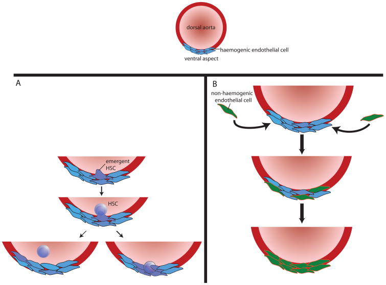
    1. Adamo L, et al. Biomechanical forces promote embryonic haematopoiesis. Nature. 2009;459:1131–5. Demonstrates that shear stress increases Runx1 expression and colony-forming potential in embryonic stem cells differentiated in vitro into HSCs, and in hematopoietic precursors in the AGM region of mouse embryos. - PMC - PubMed

Publication types