. 2010 Sep;23(3):149-60.
doi: 10.1055/s-0030-1262982.

Radiographic and endoscopic diagnosis and treatment of enterocutaneous fistulas

Affiliations

Radiographic and endoscopic diagnosis and treatment of enterocutaneous fistulas

Jennifer K Lee et al. Clin Colon Rectal Surg. 2010 Sep.

Abstract

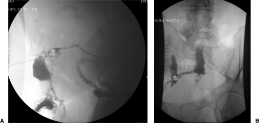
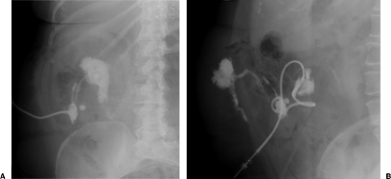
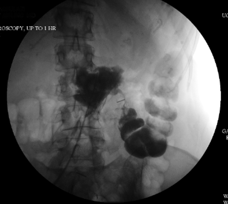
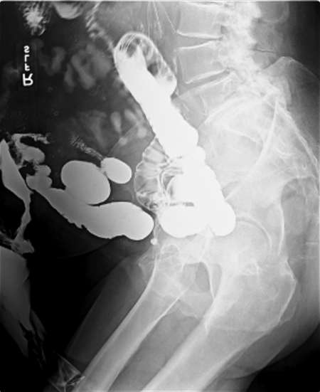
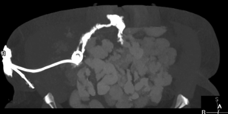
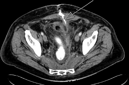
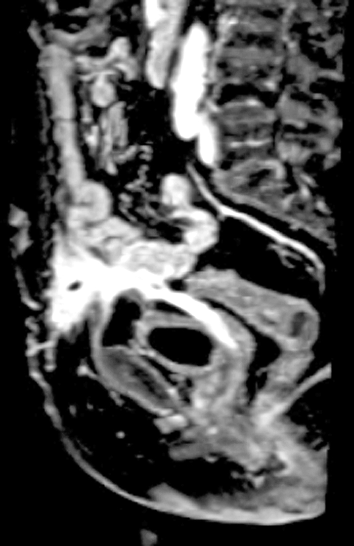
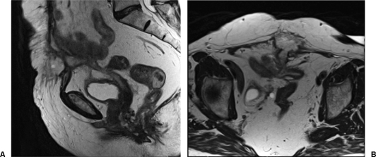
The management of enterocutaneous fistulas continues to be a challenging postoperative complication. Understanding the anatomy of the fistula optimizes its evaluation and management. Diagnostic radiology has always played an important role in this task. The use of plain radiography with contrasted studies and fistulograms is well documented in the earliest investigations of fistulas and they continue to be helpful techniques. The imaging techniques have evolved rapidly over the past 15 years with the introduction of cross-sectional imaging, ultrasound and endoscopy. The purpose of this chapter is to review both the diagnostic and therapeutic roles of fistulograms, small bowel follow-through, computed tomography, magnetic resonance imaging, ultrasound, and endoscopy in the setting of acquired enterocutaneous fistulas.

Keywords: Enterocutaneous fistula; computed tomography; fistulogram; magnetic resonance imaging; small bowel follow-through.


Figures

Figure 1
Figure 1
Two images depicting fistulograms with fluoroscopy. Note the Foley catheter balloon used to minimize backward flow of contrast and the contrast filled small bowel.
Figure 2
Figure 2
Two plain film images of a fistulogram in the same patient status postappendectomy depicting an enterocutaneous fistula to the terminal ileum.
Figure 3
Figure 3
Fluoroscopic image after percutaneous contrast administration in a patient with enterocutaneous fistula confirmed to be originating at the anastomosis of a transverse colon resection. The midline contrast collection identifies an abscess and the thin trail of contrast leads to the transverse colon. Note the surgical clip along the fistula tract.
Figure 4
Figure 4
Small bowel follow-through in a patient who had undergone multiple operations for an abdominal hernia. Note the contrast delineating the fistula tract to the left of the image and filling the colostomy bag.
Figure 5
Figure 5
Computed tomography for the same patient described in Figure 2.
Figure 6
Figure 6
Computed tomography for a patient who developed an enterocutaneous fistula after an ileostomy takedown. The arrow points to the tract that leads to the small bowel.
Figure 7
Figure 7
Magnetic resonance imaging (T1 weighted, sagittal view) in a patient who developed an enterocutaneous fistula (ECF) after Hartmann's procedure for diverticulitis. The ECF is noted at the level of the skin, leading to an abscess and then leading to the pouch and the vaginal pouch. (The patient was also status posthysterectomy.)
Figure 8
Figure 8
Magnetic resonance imaging (T2-weighted, sagittal view to left and axial view to right) for the same patient as Figure 7. The large fistula tract is seen with communication to the sigmoid pouch as well as the vaginal stump.
Figure 9
Figure 9
Cannulation of internal orifice of an enterocutaneous fistula at previous jejunostomy site.
Figure 10
Figure 10
Injection of fibrin glue.
Figure 11
Figure 11
Control of endoscopically placed Surgisis (Cook Biotech, West Lafayette, IN) at external orifice.
Figure 12
Figure 12
Stent placed into distal colostomy fistula.

References

    1. Rankin F W, Bargen J A, Buie L A. The Colon, Rectum, and Anus. Philadelphia: Saunders; 1932. p. 346.
    1. Lewis D, Penick R M. Fecal fistula. Internat Clin. 1933;1:111.
    1. Ransom H K, Coller F A. Intestinal fistula. J Mich State Med S. 1935;34:281.
    1. Frazier C H. The use of iodized rape-seed oil (campiodol) for rontgenographic exploration. Ann Surg. 1929;89(6):801–812. - PMC - PubMed
    1. Ginzburg L, Benjamin E W. Lipiodol studies of postoperative biliary fistulae. Ann Surg. 1930;91(2):233–241. - PMC - PubMed