. 2010 Sep;6(2):214-8.
doi: 10.1007/s11420-010-9170-3. Epub 2010 Jul 7.

Traumatic Lateral Plantar Artery Pseudoaneurysm and the Use of Time-Resolved MR Angiography

Affiliations

Traumatic Lateral Plantar Artery Pseudoaneurysm and the Use of Time-Resolved MR Angiography

Akira M Murakami et al. HSS J. 2010 Sep.

Abstract

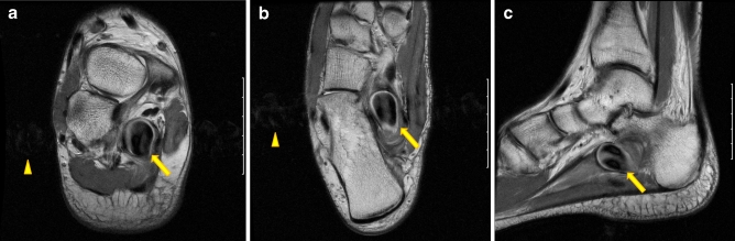
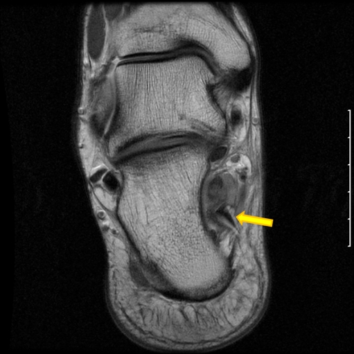
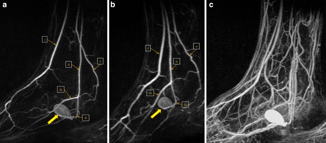
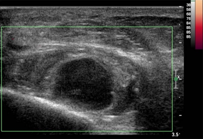
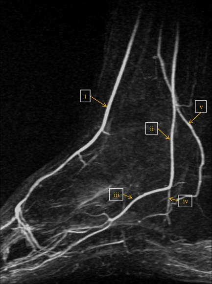
Vascular injury resulting in pseudoaneurysm formation in the plantar aspect of the foot is an uncommon injury after trauma. Such injuries are more often reported in the lateral plantar artery rather than the medial plantar artery, most likely because of its more superficial location. Traditional modalities in diagnosis have included ultrasound and digital subtraction angiography. We present a case of traumatic pseudoaneurysm of the lateral plantar artery following a foot laceration. Diagnosis was made by the use of high-resolution, time-resolved contrast-enhanced 3D magnetic resonance angiography, also referred to as "TRICKS" (time-resolved imaging of contrast kinetics). This technique provided high spatial resolution for the arterial anatomy as well as temporal resolution which allowed better delineation of the hemodynamic characteristics of the pseudoaneurysm.

Electronic supplementary material: The online version of this article (doi:10.1007/s11420-010-9170-3) contains supplementary material, which is available to authorized users.

Keywords: TRICKS; magnetic resonance angiography; plantar artery pseudoaneurysm; time-resolved imaging of contrast kinetics.


Figures

Fig. 1
Fig. 1
Oblique coronal (a), axial (b), and sagittal (c) fast spin echo MR images of the ankle demonstrate a 3.3 × 3 × 2 cm low signal intensity mass (arrows) along the medial aspect of the calcaneal body, mostly within the substance of the quadratus plantae muscle. The mass exhibits flow-related artifacts, including signal void from turbulent flow and pulsation artifact in the phase-encoding direction (arrow head), suspicious for a pseudoaneurysm
Fig. 2
Fig. 2
Oblique coronal fast spin echo MR image of the ankle demonstrates a triangular 1.0 × 0.5 cm low signal intensity foreign body (arrow) medial to the base of the lesion, later confirmed to represent a retained glass fragment
Fig. 3
Fig. 3
Arterial phase (a, b) and venous phase (c) time-resolved contrast-enhanced 3D MR angiography demonstrates rapid contrast filling and delayed washout of a pseudoaneurysm (arrow) of the lateral plantar artery (iv), 3 cm distal to the bifurcation of the posterior tibial artery (ii). The lateral plantar artery continued distally beyond the pseudoaneurysm to complete the plantar arch. (i) Anterior tibial artery, (ii) posterior tibial artery, (iii) medial plantar artery, (iv) lateral plantar artery, and (v) distal fibular arteries [21]
Fig. 4
Fig. 4
Power Doppler ultrasound at 3 weeks follow-up demonstrates no blood flow within the hypoechoic pseudoaneurysm, indicating complete thrombosis
Fig. 5
Fig. 5
Sagittal fast spin MR image of the ankle at 2 months follow-up demonstrates high signal intensity within the pseudoaneurysm (arrow), without flow-related signal voids or pulsation artifact, indicating complete thrombosis
Fig. 6
Fig. 6
TRICKS MR angiography at 2 months follow-up demonstrates diminished flow within the lateral plantar artery and the complete lack of contrast filling of the pseudoaneurysm, indicating complete thrombosis. (i) Anterior tibial artery, (ii) posterior tibial artery, (iii) medial plantar artery, (iv) lateral plantar artery, and (v) distal fibular arteries

Similar articles

Cited by

References

    1. Agarwal M, Harkless L, Hagino RT, Toursarkissian B. Lateral plantar artery aneurysm: a case report. J Am Podiatr Med Assoc. 2007;97(6):480–2. - PubMed
    1. Nierenberg G, Hoffman A, Engel A, Stein H. Pseudoaneurysm with an arteriovenous fistula of the tibial vessels after plantar fasciotomy: a case report. Foot Ankle Int. 1997;18(8):524–5. - PubMed
    1. Gentile AT, Zizzo CJ, Dahukey A, Berman SS. Traumatic pseudoaneurysm of the lateral plantar artery after endoscopic plantar fasciotomy. Foot Ankle Int. 1997;18(12):821–2. - PubMed
    1. Ptaszek AJ, Aminian A, Schneider JR, Milos S. Lateral plantar artery pseudoaneurysm after calcaneal osteotomy: a case report. Foot Ankle Int. 2006;27(2):141–3. - PubMed
    1. Thornton BP, Minion DJ, Quick R, Vasconez HC, Endean ED. Pseudoaneurysm of the lateral plantar artery after foot laceration. J Vasc Surg. 2003;37(3):672–5. doi: 10.1067/mva.2003.31. - DOI - PubMed