Case Reports
. 2010 Sep;6(2):228-34.
doi: 10.1007/s11420-010-9159-y. Epub 2010 Mar 9.

Traumatic osteochondral injury of the femoral head treated by mosaicplasty: a report of two cases

Case Reports

Traumatic osteochondral injury of the femoral head treated by mosaicplasty: a report of two cases

Denis Nam et al. HSS J. 2010 Sep.

Abstract

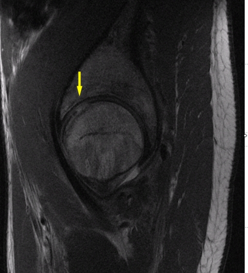
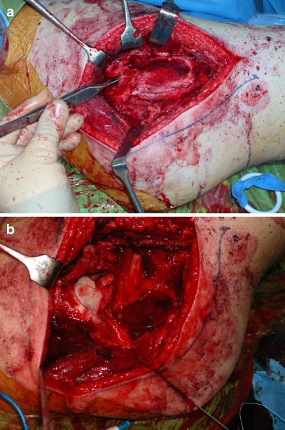
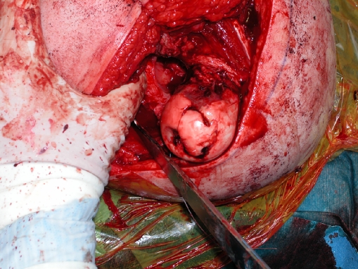
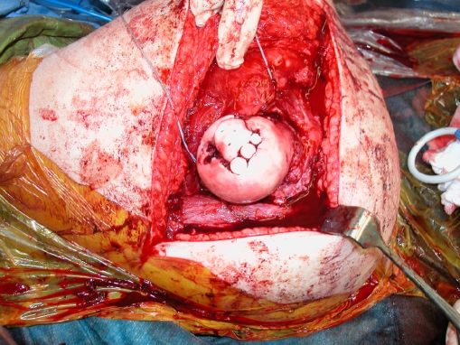
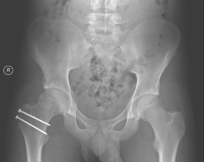
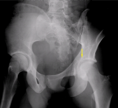
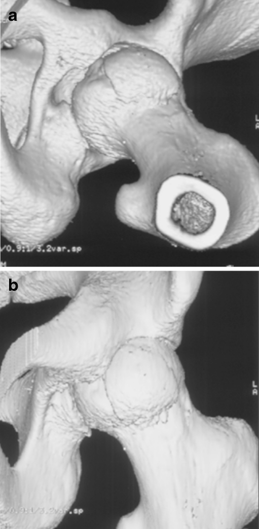
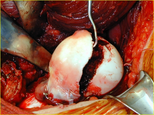
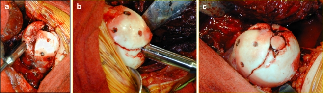
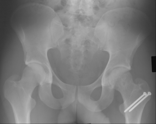
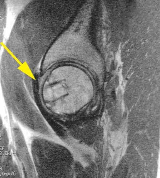
The increased risk of symptomatic progression towards osteoarthritis after chondral damage has led to the development of multiple treatment options for cartilage repair. These procedures have evolved from arthroscopic lavage and debridement, to marrow stimulation techniques, and more recently, to osteochondral autograft and allograft transplants, and autogenous chondrocyte implantation. The success of mosaicplasty procedures in the knee has led to its application to other surfaces, including the talus, tibial plateau, patella, and humeral capitellum. In this report, we present two cases of a chondral defect to the femoral head after a traumatic hip dislocation, treated with an osteochondral autograft (OATS) from the ipsilateral knee, and the inferior femoral head, respectively, combined with a surgical dislocation of the hip. At greater than 1 year and greater than 5 years of follow-up, MRI studies have demonstrated good autograft incorporation with maintenance of articular surface conformity, and both patients clinically continue to have no pain and full active range of motion of their respective hips. In our opinion, treatment of osteochondral defects in the femoral head surface using a surgical dislocation combined with an OATS procedure is a promising approach, as full exposure of the femoral head can be obtained while preserving its vasculature, thus enabling adequate restoration of the articular cartilage surface.

Keywords: femoral head; hip dislocation; mosaicplasty; osteochondral autograft transplant; osteochondral defect; osteochondral injury.


Figures

Fig. 1
Fig. 1
A preoperative AP radiograph of the pelvis demonstrating no appreciable abnormality in the contour of the femoral head
Fig. 2
Fig. 2
A sagittal section of the patient’s MRI demonstrating an area of full-thickness cartilage loss in the anterior-superior weight-bearing zone of the femoral head
Fig. 3
Fig. 3
Intraoperative images demonstrating the trochanteric osteotomy (a) and exposure of the femoral head (b) prior to dislocating the femoral head anteriorly
Fig. 4
Fig. 4
An intraoperative image demonstrating cartilage impaction along with exposed subchondral bone in the anterior-superior portion of the femoral head
Fig. 5
Fig. 5
An intraoperative image of the femoral head after placement of the donor osteochondral plugs and three SmartNails
Fig. 6
Fig. 6
A postoperative AP radiograph of the patient’s pelvis demonstrating an anatomic contour of the femoral head and well-preserved joint space
Fig. 7
Fig. 7
A preoperative internal (obturator) oblique view of the left hip, demonstrating an intra-articular bony fragment in the left hip
Fig. 8
Fig. 8
Preoperative 3D reconstructed images demonstrating a vertically oriented femoral head fracture extending above and below the fovea in the coronal plane
Fig. 9
Fig. 9
An intraoperative image demonstrating a large, Pipkin type II femoral head osteochondral fracture
Fig. 10
Fig. 10
An intraoperative image demonstrating placement of a 2.7-mm PLA Bionix (Bionix Incorporation, Toledo, OH, USA) screw for fixation of the osteochondral fragment. Three screws were placed in total
Fig. 11
Fig. 11
Intraoperative images demonstrating the zone of injury at the anterior-superior weight-bearing dome, and harvesting of the osteochondral autograft plug from the inferior aspect of the femoral head (a, b). An image demonstrating a smooth articular contour of the femoral head after impaction of the donor plug (c)
Fig. 12
Fig. 12
An AP pelvis radiograph at 1 year post-operatively demonstrating good preservation of the joint space and anatomic contour of the femoral head
Fig. 13
Fig. 13
Sagittal MRI image at 1 year post-operatively, demonstrating a well-incorporated autograft plug with no focal full-thickness defect and consistent cartilaginous signal and architecture throughout the femoral head

Similar articles

Cited by

References

    1. Alford JW, Cole BJ. Cartilage restoration, part 2: Techniques, outcomes, and future directions. Am J Sports Med. 2005;33:443–460. doi: 10.1177/0363546505274578. - DOI - PubMed
    1. Brittberg M, Lindahl A, Nilsson A, Ohlsson C, Isaksson O, Peterson L. Treatment of deep cartilage defects in the knee with autologous chondrocyte transplantation. N Engl J Med. 1994;331:889–895. doi: 10.1056/NEJM199410063311401. - DOI - PubMed
    1. Steadman JR, Rodkey WG, Briggs KK. Microfracture to treat full-thickness chondral defects: Surgical technique, rehabilitation, and outcomes. J Knee Surg. 2002;15:170–176. - PubMed
    1. Alford JW, Cole BJ. Cartilage restoration, part 1: Basic science, historical perspective, patient evaluation, and treatment options. Am J Sports Med. 2005;33:295–306. doi: 10.1177/0363546504273510. - DOI - PubMed
    1. Buckwalter JA, Mow VC, Ratcliffe A. Restoration of injured or degenerated articular cartilage. J Am Acad Orthop Surg. 1994;2:192–201. - PubMed

Publication types