

A ventricular thrombus mimicking a tumour
- PMID: 21886661
- PMCID: PMC3028419
- DOI: 10.1136/bcr.06.2009.1944
A ventricular thrombus mimicking a tumour
Abstract
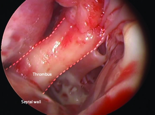
Primary cardiac tumours are a rare occurrence in clinical practice. Mural or pedunculated thrombi are other infrequent findings. These are usually associated with underlying heart disease, present in the left atrium or occupying aneurysms in the ventricular wall, usually the apex. The case of a 33-year-old woman with a pedunculated cardiac mass not having these latter characteristics is reported. She had experienced dyspnoea and lower extremity oedema for 3 years on referral. Echocardiography revealed a mass emerging from the interventricular septum, and a myxoma was suspected. Heart surgery was performed and the findings were a thrombus and large extent of septal and apical mural fibrosis. An endoaneurysmorraphy was performed with exclusion of fibrotic walls from the ventricular cavity.
Figures


References
-
- van Dantzig JM, Delemarre BJ, Bot H, et al. Left ventricular thrombus in acute myocardial infarction. Eur Heart J 1996; 17: 1640–5 - PubMed
-
- Waller BF, Rohr TM, McLaughlin T, et al. Intracardiac thrombi: frequency, location, etiology, and complications: a morphologic review - part V. Clin Cardiol 1995; 18: 731–4 - PubMed
-
- Burke A, Jeudy J, Jr, Virmani R.Cardiac tumors.: Topol EJ, Califf RM, Isner J, et al., eds. Textbook of cardiovascular medicine. 3rd edn Philadelphia, Pennsylvania, USA: Lippincott-Raven, 2006
-
- Maraj S, Pressman GS, Figueredo VM. Primary cardiac tumors. Int J Cardiol 2009; 133: 152–6 - PubMed
LinkOut - more resources
Full Text Sources
Miscellaneous