

Trichoscopy (hair and scalp videodermoscopy) in the healthy female. Method standardization and norms for measurable parameters
- PMID: 21886722
- PMCID: PMC3157785
- DOI: 10.3315/jdcr.2008.1021
Trichoscopy (hair and scalp videodermoscopy) in the healthy female. Method standardization and norms for measurable parameters
Abstract
Background: Trichoscopy is a newly developed method of hair image analysis, based on videodermoscopy of hair and scalp.
Objective: The aim of the study was to establish normal values and set the standard for trichoscopy in female population.
Patients and methods: A total of 60 healthy females with no symptoms of hair or scalp diseases in anamnesis, upon clinical examination and in classic hair diagnostic techniques were included into the study. Mean age of these females was 36.5 (19-64) years. Trichoscopy was performed with the use of Fotofinder II. In all patients trichoscopy was performed in four locations (frontal area, occipital area, left and right temporal area). Hair and perifollicular area were evaluated. Measurements were performed with the application of the MoleAnalyzer software.
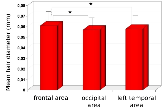
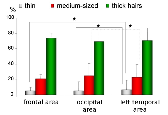
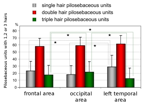
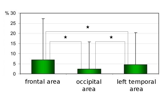
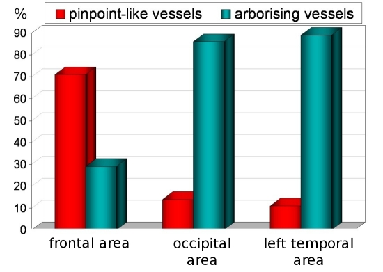
Results: Mean hair thickness was 0.061mm±0.008mm in frontal area vs. 0.057mm±0.007mm in occiput (p<0,001) and vs. 0.058mm±0.008mm in left temporal area and 0.059mm±0.008mm in right temporal area (p>0.005). The percentage of thin hairs (below 0.03mm) was 5%±4.3 in frontal area vs. 5.5%±4.8 in occiput vs. 6.4%±5.7 in right temporal area. The highest proportion of single-hair pilosebaceous units was observed in the temporal areas (29.1±16.2 vs. 23.2±13.5 in frontal and 18.4±12.1 in occipital areas; p<0.005). Based on study results, the norms for parameters measured in trichoscopy were assessed: mean hair thickness bigger than 0,053mm in frontal area and bigger than 0,050mm in others; percentage of thin hairs should be less than 10% in frontal and occipital area and less than 13% in temporal areas. The percentage of pilosebaceous units with single hair should be less than 35% in frontal area, 30% in occiput and 40% in temporal areas. Yellow dots were seen sporadically and they shouldn't be in a higher number than 3 in 4 fields of vision with 70-fold magnification in frontal area and only 1 in others. Perifollicular discoloration should be lower than 25% for frontal area, lower than 15% in occiput and 20% for temporal areas.
Conclusion: A standard procedure to perform trichoscopy (hair and scalp videodermoscopy) for diagnostic purposes was developed. Norms of measurable parameters were established for the population of adult white females.
Keywords: alopecia; dermoscopy; hair; trichoscopy; videodermoscopy; women.
Figures
References
-
- Olszewska M, Rudnicka L, Rakowska A, Kowalska-Oledzka E, Slowinska M. Trichoscopy. Arch Dermatol. 2008;144:1007. - PubMed
-
- Rudnicka L, Olszewska M, Majsterek M, Czuwara J, Slowinska M. Presence and future of dermoscopy. Expert Rev Dermatol. 2006;1:769–772.
-
- Lacarrubba F, Dall'Oglio F, Rita Nasca M, Micali G. Videodermatoscopy enhances diagnostic capability in some forms of hair loss. Am J Clin Dermatol. 2004;5:205–208. - PubMed
-
- Ross EK, Vincenzi C, Tosti A. Videodermoscopy in the evaluation of hair and scalp disorders. J Am Acad Dermatol. 2006;55:799–806. - PubMed
-
- Slowinska M, Rudnicka L, Schwartz RA, Kowalska-Oledzka E, Rakowska A, Sicinska J, Lukomska M, Olszewska M, Szymanska E. Comma hairs: a dermatoscopic marker for tinea capitis: a rapid diagnostic method. J Am Acad Dermatol. 2008;59 (5 suppl):S77–79. - PubMed
LinkOut - more resources
Full Text Sources
Medical