. 2011;6(8):e22968.
doi: 10.1371/journal.pone.0022968. Epub 2011 Aug 23.

Fukutin-related protein resides in the Golgi cisternae of skeletal muscle fibres and forms disulfide-linked homodimers via an N-terminal interaction

Affiliations

Fukutin-related protein resides in the Golgi cisternae of skeletal muscle fibres and forms disulfide-linked homodimers via an N-terminal interaction

Maisoon Alhamidi et al. PLoS One. 2011.

Abstract

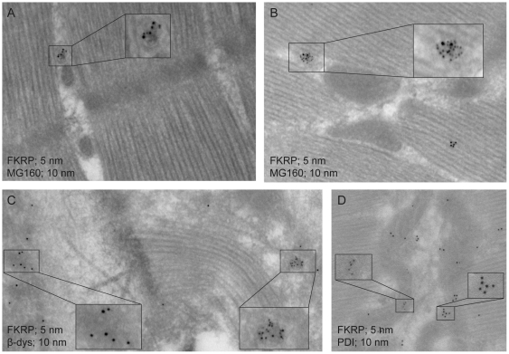
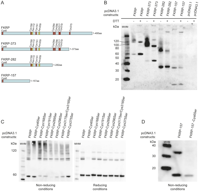
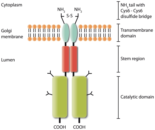
Limb-Girdle Muscular Dystrophy type 2I (LGMD2I) is an inheritable autosomal, recessive disorder caused by mutations in the FuKutin-Related Protein (FKRP) gene (FKRP) located on chromosome 19 (19q13.3). Mutations in FKRP are also associated with Congenital Muscular Dystrophy (MDC1C), Walker-Warburg Syndrome (WWS) and Muscle Eye Brain disease (MEB). These four disorders share in common an incomplete/aberrant O-glycosylation of the membrane/extracellular matrix (ECM) protein α-dystroglycan. However, further knowledge on the FKRP structure and biological function is lacking, and its intracellular location is controversial. Based on immunogold electron microscopy of human skeletal muscle sections we demonstrate that FKRP co-localises with the middle-to-trans-Golgi marker MG160, between the myofibrils in human rectus femoris muscle fibres. Chemical cross-linking experiments followed by pairwise yeast 2-hybrid experiments, and co-immune precipitation, demonstrate that FKRP can exist as homodimers as well as in large multimeric protein complexes when expressed in cell culture. The FKRP homodimer is kept together by a disulfide bridge provided by the most N-terminal cysteine, Cys6. FKRP contains N-glycan of high mannose and/or hybrid type; however, FKRP N-glycosylation is not required for FKRP homodimer or multimer formation. We propose a model for FKRP which is consistent with that of a Golgi resident type II transmembrane protein.


Conflict of interest statement

Competing Interests: The authors have declared that no competing interests exist.

Figures

Figure 1
Figure 1. FKRP subcellular localisation in skeletal muscle, determined by double EM immunogold labelling.
FKRP was detected with a mixture of FKRP207 and FKRP208 primary antibodies followed by labelling with goat anti-rabbit antibody, conjugated with 5 nm gold particles (A, B, C and D). Subcellular marker antibodies were, anti MG160 (Golgi) (A, B), anti ß-dys (sarcolemma) (C) and anti PDI (endoplasmic reticulum) (D). Secondary antibody for detection of subcellular marker proteins was goat anti-mouse antibody, conjugated with 10 nm gold particles. Selected areas (framed) were enlarged by 200%.
Figure 2
Figure 2. Recombinant FKRP forms multimers in mammalian cells.
COS-7 and BHK-21 cells were transfected with pcDNA3.1-FKRP or empty vector (pcDNA3.1). Forty-eight hrs after transfection cells were solubilised in lysis buffer with protease inhibitor. A) The cleared COS-7 lysate was either kept untreated (lane 1) or reduced with 400 mM DTT at RT for 30 min (lane 2) or Sample Reducing agent at 99°C for 5 min (lanes 3 and 4) and subsequently subjected to (4–12%) SDS-PAGE and Western blot analysis. Lanes 1–3: lysates from pcDNA3.1-FKRP transfections. Lane 4: lysate from pcDNA3.1 transfection serving as negative control. B) A cleared BHK-21 lysate was treated with 10 mM EGS at RT for 30 min and quenched with 35 mM Tris, pH 7.5 for 15 min. The EGS treated sample and the accompanying untreated control sample were reduced with Sample Reducing agent at 99°C for 5 min and subjected to SDS-PAGE and Western blot analysis. In the above experiments FKRP was detected using FKRP207 antibodies. MWM is molecular weight marker.
Figure 3
Figure 3. FKRP self-interaction as demonstrated by pair wise Y2H analysis and CO-IP experiments.
A) Diploid yeast cells containing both bait construct, pB27-FKRP (N-LexA-FKRP32-494-C), and prey construct, pP7-FKRP (N-GAL4-FKRP32-494-C), were obtained by mating and spotted, at the dilutions indicated, onto non-selective media lacking Trp and Leu (left panel) and selective media lacking Trp, Leu and His (right panel). Negative controls contained empty bait and pray vectors, pB27 and pP7, or pB27 and pP7 in combination with prey and bait constructs, respectively. Positive control (C+) contained human SMAD3 as bait (GI:5174512) and Human SMURF1 as prey (GI:31317291) as explained in Materials and Methods. B) Anti-Myc antibody was employed to precipitate FKRP-Myc fusion proteins from COS-7 cell lysates. The lysates were prepared by pcDNA3.1-FKRP-Myc/pcDNA3.1-FKRP-HA co-transfection (Cotr) or solo transfections of pcDNA3.1-FKRP-Myc and pcDNA3.1-FKRP-HA. An additional sample was prepared by mixing pcDNA3.1-FKRP-Myc and pcDNA3.1-FKRP-HA lysates followed by incubation at RT for 30 min (Mix). All cell lysates used for Co-IP were prepared in the presence of 5 mM NEM. Lysates from pcDNA3.1 and pcDNA3.1-FKRP-HA solo transfections served as negative controls whereas lysates from pcDNA3.1-Myc transfected cells served as positive control for anti-Myc based immune precipitation. Input samples and precipitates were subjected to (4–12%) SDS-PAGE, under reducing conditions, followed by Western blot analysis. On separate blots FKRP-Myc and FKRP-HA were detected with anti-Myc and anti-HA antibodies, respectively. To asses the stringency of the of the Co-IP experiment, one of the blots (anti-HA) was stripped and assayed for endogenous MAPK with anti-MAPK antibody (lower panel).
Figure 4
Figure 4. FKRP dimer and multimer formation is not dependent on N-glycosylation.
Cells were transfected with pcDNA 3.1-FKRP. Forty-eight hrs after transfection cells were solubilised in lysis buffer with protease inhibitor. A) A cleared BHK-21 lysate was treated with either PNGase F or Endo H as explained in Materials and Methods and subjected to (4–12%) SDS-PAGE under reducing conditions, followed by Western blot analysis. An untreated sample served as control. B) COS-7 cells were transfected with mutant pcDNA3.1-FKRP constructs, in which either one or both asparagines (Asn) involved in N-glycosylation had been replaced with glutamine (Gln). The lysates were prepared in the presence of 5 mM NEM and subjected to either non-reducing (left panel) or reducing SDS-PAGE (330 mM DTT, RT for 30 min) (right panel), followed by Western blot analysis. Antibody FKRP207 was used for the detection of FKRP in these experiments.
Figure 5
Figure 5. FKRP homodimer formation depends on a Cys6-Cys6 disulfide bridge.
A) Schematic presentation FKRP C-terminal deletion constructs. FKRP-373, FKRP-282 and FKRP-157 denote the length (aa) of the mutant construct. Red vertical bars show Cys (C) positions whereas yellow vertical bars indicate the positions of putative N-glycosylation sites Asn-X-Thr/Ser (N-X-T/S). In the following experiments COS-7 cells were transfected with various pcDNA3.1 constructs and cleared lysates were prepared 48 hrs post transfection in the presence of 5 mM NEM. In all these experiments FKRP was detected with primary antibody FKRP207. B) Cleared lysates from FKRP C-terminal deletion mutants were either left untreated or subjected to reduction (400 mM DTT, at RT for 30 min), followed by (4–12%) SDS-PAGE and Western blot analysis. C) Cleared lysates from FKRP Cys→Ser substitution mutants were either left untreated (left panel) or subjected to reduction (400 mM DTT, at RT, for 30 min) (right panel), followed by (4–12%) SDS-PAGE and Western blot analysis. Differences in migration between monomeric forms as well as between dimeric forms (5C) likely represent different FKRP conformations resolved by SDS-PAGE. Such conformational differences might be induced by Cys→Ser mutations as some of the mutant Cys residues must be expected to be involved in intra-molecular disulfide bridges. D) COS-7 lysates from deletion construct FKRP-157, and the Cys6Ser mutant thereof, were subjected to SDS-PAGE under non-reducing conditions, followed by Western blot analysis.
Figure 6
Figure 6. Proposed model of the Golgi resident FKRP dimer; a putative glycosyltransferase.
According to the type II transmembrane glycosyltransferase model, the architecture comprises a globular catalytic domain, a stem region, a single pass transmembrane domain and a cytoplasmic N-terminal tail , , . Based on the present work we suggest that FKRP forms homodimers via an interaction interface that extends through the stem region. The interaction is stabilised by a Cys6-Cys6 disulfide bridge in the N-terminal cytoplasmic tail resulting in a covalently connected FKRP dimer with two-fold symmetry. The catalytic domain is likely to interact with other proteins forming large multimeric structures (not depicted). -S-S-; disulfide bridge, Y; N-glycan of high mannose and/or hybrid type.

References

    1. Martin PT. Mechanisms of disease: congenital muscular dystrophies- glycosylation takes center stage. Nat Clin Pract Neurol. 2006;2:222–230. - PMC - PubMed
    1. Kanagawa M, Toda T. The genetic and molecular basis of muscular dystrophy: roles of cell-matrix linkage in the pathogenesis. J Hum Genet. 2006;51:915–926. - PubMed
    1. Barresi R, Campbell KP. Dystroglycan: from biosynthesis to pathogenesis of human disease. J Cell Sci. 2006;119:199–207. - PubMed
    1. Henry MD, Campbell KP. Dystroglycan inside and out. Curr Opin Cell Biol. 1999;11:602–607. - PubMed
    1. Ervasti JM, Campbell KP. Membrane organization of the dystrophin- glycoprotein complex. Cell. 1991;66:1121–1131. - PubMed

Publication types

MeSH terms