. 2011;6(8):e23344.
doi: 10.1371/journal.pone.0023344. Epub 2011 Aug 23.

Monoclonal antibodies recognizing the non-tandem repeat regions of the human mucin MUC4 in pancreatic cancer

Affiliations

Monoclonal antibodies recognizing the non-tandem repeat regions of the human mucin MUC4 in pancreatic cancer

Maneesh Jain et al. PLoS One. 2011.

Abstract

The MUC4 mucin is a high molecular weight, membrane-bound, and highly glycosylated protein. It is a multi-domain protein that is putatively cleaved into a large mucin-like subunit (MUC4α) and a C-terminal growth-factor like subunit (MUC4β). MUC4 plays critical roles in physiological and pathological conditions and is aberrantly overexpressed in several cancers, including those of the pancreas, cervix, breast and lung. It is also a potential biomarker for the diagnosis, prognosis and progression of several malignancies. Further, MUC4 plays diverse functional roles in cancer initiation and progression as evident from its involvement in oncogenic transformation, proliferation, inhibition of apoptosis, motility and invasion, and resistance to chemotherapy in human cancer cells. We have previously generated a monoclonal antibody 8G7, which is directed against the TR region of MUC4, and has been extensively used to study the expression of MUC4 in several malignancies. Here, we describe the generation of anti-MUC4 antibodies directed against the non-TR regions of MUC4. Recombinant glutathione-S-transferase (GST)-fused MUC4α fragments, both upstream (MUC4α-N-Ter) and downstream (MUC4α-C-Ter) of the TR domain, were used as immunogens to immunize BALB/c mice. Following cell fusion, hybridomas were screened using the aforementioned recombinant proteins ad lysates from human pancreatic cell lines. Three anti MUC4α-N-Ter and one anti-MUC4α-C-Ter antibodies were characterized by several inmmunoassays including enzyme-linked immunosorbent assay (ELISA), immunoblotting, immunofluorescene, flow cytometry and immunoprecipitation using MUC4 expressing human pancreatic cancer cell lines. The antibodies also reacted with the MUC4 in human pancreatic tumor sections in immunohistochemical analysis. The new domain-specific anti-MUC4 antibodies will serve as important reagents to study the structure-function relationship of MUC4 domains and for the development of MUC4-based diagnostics and therapeutics.


Conflict of interest statement

Competing Interests: The authors have declared that no competing interests exist.

Figures

Figure 1
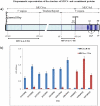
Figure 1. Schematic structure of the recombinant MUC4 domains and reactivity of various anti-MUC4 antibodies.
a) Schematic structure of MUC4 and recombinant proteins used in the study. MUC4 is putatively cleaved at the GDPH site to generate an N-terminal mucin-type subunit MUC4-α and a C-terminal growth factor-type subunit MUC4-β. Important domains of MUC4 are marked. Recombinant domains of MUC4- α corresponding to the fragments upstream and downstream of the tandem-repeat (TR) domain were cloned and expressed as described in Materials and Methods and termed MUC4-α-N-ter and MUC4-α-C-Ter, respectively. The nucleotide numbers corresponding to the boundaries of the recombinant domains are marked and are described in Moniaux et al. and Choudhury et al (Ref 1 and 24, respectively) according to the original numbering. Cys-cystein-rich domain EGF-epidermal growth factor-like domain; TM-transmembrane domain; CT-cytoplasmic tail. b) ELISA showing the reactivity of anti-MUC4 MAbs to recombinant immunogens. The indicated MAbs were incubated with the 2.5 µg/ml of GST-tagged N-terminal and tandem repeat recombinant domains of MUC4. The specificities were also tested against the MUC4 TR peptide, GST and a non-specific control protein bovine serum albumin and the antibodies exhibited negative reactivity against these antigens. The assay also included a non-specific isotype matched control K2G6.
Figure 2
Figure 2. Comparative immunoblot analysis for MUC4 expression in various pancreatic cancer cell lines using various antibodies.
A total of 20 µg of protein from cell extracts was resolved by electrophoresis on a 2% SDS-agarose gel, transferred to PVDF membrane, and incubated with 2 µg/ml of MAbs 2175 (a), 2214(b), 2382 (c) or 1 µg/ml of anti-MUC4 TR Mab 8G7(d). The membrane was then probed with horseradish peroxidase-labeled goat anti-mouse immunoglobulin. The signal was detected using an ECL reagent kit. The position of the detected bands is indicated by arrows. For loading control, immunoblot for the detection of β-actin (inset a) was done on lysates of respective cells resolved on 10% SDS-PAGE.
Figure 3
Figure 3. Immunofluorescence of MUC4 in CD18/HPAF cells with various anti-MUC4 MAbs.
Cells were grown at low density on sterilized cover-slides, fixed in ice-cold methanol at −20°C and were incubated with 10 µg/ml non-TR MAbs of 2214, 2175, 2382 and 2106, or 2 µg/ml of anti-MUC4 TR MAb 8G7 (Control) and detected using FITC conjugated secondary antibody. Anti-KLH antibody K2G6 was used as an isotype control. Cells were mounted on glass slides using anti-fade Vectashield mounting medium and observed under a ZEISS confocal laser scanning microscope (magnification, ×630).
Figure 4
Figure 4. Cell-surface binding analysis of anti-MUC4 antibodies.
Cells were harvested non-enzymatically, fixed with paraformaldehyde and incubated with the indicated antibodies. Following incubation with secondary antibody, cells were analyzed using BD FACSCalibur. The mean fluorescence intensities (MFI) values obtained with each antibody is indicated in parantheses.
Figure 5
Figure 5. Immunoprecipitation of MUC4 using various MAbs to MUC4.
Protein lysates from the MUC4-expressing CD18/HPAF cells were immunoprecipitated using 5 µg/ml of 8G7 (Tandem repeat MAb), 2382, 2214 and 2175 (Non-tandem repeat MAbs) and K2G6 (Isotype matched control MAb) and were immunoblotted using MAbs 8G7 and 2214 as described in Materials and Methods.
Figure 6
Figure 6. Immunoperoxidase staining for MUC4 in pancreatic cancer tissues using non-TR MAbs.
Paraffin sections were incubated with the indicated test and control antibodies and binding was detected using VECTOR Universal staining Kit. MAb 8G7 was used at a concentration of 2 µg/ml, while all other antibodies were used at a concentration of 10 µg/ml.

Similar articles

Cited by

References

    1. Moniaux N, Nollet S, Porchet N, Degand P, Laine A, et al. Complete sequence of the human mucin MUC4: a putative cell membrane-associated mucin. Biochem J. 1999;338:325–333. - PMC - PubMed
    1. Chaturvedi P, Singh AP, Batra SK. Structure, evolution, and biology of the MUC4 mucin. FASEB J. 2008;22:966–981. - PMC - PubMed
    1. Sheng Z, Wu K, Carraway KL, Fregien N. Molecular cloning of the transmembrane component of the 13762 mammary adenocarcinoma sialomucin complex. A new member of the epidermal growth factor superfamily. J Biol Chem. 1992;267:16341–16346. - PubMed
    1. Wu K, Fregien N, Carraway KL. Molecular cloning and sequencing of the mucin subunit of a heterodimeric, bifunctional cell surface glycoprotein complex of ascites rat mammary adenocarcinoma cells. J Biol Chem. 1994;269:11950–11955. - PubMed
    1. Copin MC, Devisme L, Buisine MP, Marquette CH, Wurtz A, et al. From normal respiratory mucosa to epidermoid carcinoma: expression of human mucin genes. Int J Cancer. 2000;86:162–168. - PubMed

Publication types