. 2011;6(8):e23530.
doi: 10.1371/journal.pone.0023530. Epub 2011 Aug 19.

Modulation of syndecan-1 shedding after hemorrhagic shock and resuscitation

Affiliations

Modulation of syndecan-1 shedding after hemorrhagic shock and resuscitation

Ricky J Haywood-Watson et al. PLoS One. 2011.

Abstract

The early use of fresh frozen plasma as a resuscitative agent after hemorrhagic shock has been associated with improved survival, but the mechanism of protection is unknown. Hemorrhagic shock causes endothelial cell dysfunction and we hypothesized that fresh frozen plasma would restore endothelial integrity and reduce syndecan-1 shedding after hemorrhagic shock. A prospective, observational study in severely injured patients in hemorrhagic shock demonstrated significantly elevated levels of syndecan-1 (554±93 ng/ml) after injury, which decreased with resuscitation (187±36 ng/ml) but was elevated compared to normal donors (27±1 ng/ml). Three pro-inflammatory cytokines, interferon-γ, fractalkine, and interleukin-1β, negatively correlated while one anti-inflammatory cytokine, IL-10, positively correlated with shed syndecan-1. These cytokines all play an important role in maintaining endothelial integrity. An in vitro model of endothelial injury then specifically examined endothelial permeability after treatment with fresh frozen plasma orlactated Ringers. Shock or endothelial injury disrupted junctional integrity and increased permeability, which was improved with fresh frozen plasma, but not lactated Ringers. Changes in endothelial cell permeability correlated with syndecan-1 shedding. These data suggest that plasma based resuscitation preserved endothelial syndecan-1 and maintained endothelial integrity, and may help to explain the protective effects of fresh frozen plasma after hemorrhagic shock.


Conflict of interest statement

Competing Interests: The authors have declared that no competing interests exist.

Figures

Figure 1
Figure 1. Syndecan-1 is shed following hemorrhagic shock.
Plasma syndecan-1 was measured in patients presenting in hemorrhagic shock. The mean plasma concentration is shown for controls, pre-resuscitation, and post-resuscitation time points. Syndecan-1 concentrations were markedly elevated pre-resuscitation and remained significantly elevated post-resuscitation compared to controls. Resuscitated patients reached the ICU an average of 7±0.75 hours after arrival in the emergency room. Results are presented as mean ± SEM, means notated with letters indicate statistical differences between groups.
Figure 2
Figure 2. Cytokines correlate with syndecan-1 shedding.
Scatter plots, with linear regression lines, of the cytokines identified to correlate with syndecan-1 are shown. Fractalkine, IFN-γ, and IL-1β were negatively correlated with syndecan-1. IL-10, on the other hand was positively correlated. Symbols: + normal donors, • pre-resuscitation, ▴post-resuscitation.
Figure 3
Figure 3. Endothelial cell permeability is decreased by FFP in an in vitro model of endothelial injury.
HUVEC's were stimulated with IL-1β and subjected to hypoxia for four hours followed by rexoygenation and incubation for an additional four hours in media alone (hypoxia/reoxygenation, HR), 5% lactated Ringers (LR), or 5% FFP, and compared to normoxia. In vitro permeability was assessed by fluoresceine-isothiocyanate [FITC] labeled dextran transport. Permeability was significantly increased by HR compared to normoxia, lessened by LR, but FFP decreased permeability to below that of normoxia. Results are presented as means ± SEM, n = 6/group. Means notated with letters indicate statistical differences between groups.
Figure 4
Figure 4. VE-cadherin immunoreactivity is enhanced by FFP in an in vitro model of endothelial injury.
HUVEC's were stimulated with IL-1β and subjected to hypoxia for four hours followed by rexoygenation and incubation for an additional four hours in media alone (hypoxia/reoxygenation, HR), 5% lactated Ringers (LR), or 5% FFP, and compared to normoxia. A. Cells were labeled with antibody to VE-cadherin and images captured with an IX71 inverted microscope. Original image magnification, ×40. B. The relative fluorescence intensity was quantified using Image J software (NIH) and reported as relative fluorescence units (RFU). Results are presented as means ± SEM, n = 3/group. VE-cadherin immunoreactivity was reduced by HR and LR compared to normoxia, but enhanced by FFP.
Figure 5
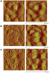
Figure 5. Endothelial surface ultrastructure is restored by FFP in an in vitro model of shock.
HUVEC's were cultured under normoxic conditions or stimulated with IL-1β and subjected to hypoxia for four hours followed by rexoygenation and incubation for an additional four hours in 5% lactated Ringers (LR), or 5% FFP and compared to normoxia. Cell surface ultrastructure was assessed using atomic force microscopy, 80 µm area scanned for all images. The deflection (left) and corresponding height (right) topographic images are shown from representative images of three separate experiments for each group. Normoxic controls (A) demonstrate no gaps between cells whereas areas of thinning between cell junctions are seen after LR (B) but reduced by FFP (C). Gaps are indicated by white lines.
Figure 6
Figure 6. Cell surface syndecan-1 is enhanced by FFP in an in vitro model of endothelial injury.
HUVEC's were stimulated with IL-1β and subjected to hypoxia for four hours followed by rexoygenation and incubation for an additional four hours in media alone (hypoxia/reoxygenation, HR), 5% lactated Ringers (LR) or5% FFP and compared to normoxia. A. Cell were labeled with antisyndecan-1 antibodies (magnification = original×200) then B. Images obtained using an Olympus 1×71 microscope with SimplePCI6 software. Original image magnification, ×20. The relative fluorescence intensity was quantified using Image J software (NIH) and reported as relative fluorescence units (RFU). Results are presented as means ± SEM, n = 6 images/group. Means notated with letters indicate statistical differences between groups. Immunostaining revealed that cell surface syndecan-1 is expressed abundantly in normoxia, significantly reduced after HR and LR, but partially restored by FFP.
Figure 7
Figure 7. Proposed model of syndecan-1 interaction with inflammatory cytokines after shock and resuscitation with FFP.
(A) Syndecan-1 (sdc-1) is a major constituent of the protective glycocalyx found on the surface of endothelial cells (ECs). (B) During hemorrhagic shock, syndecan-1 is shed from the EC surface, exposing the underlying endothelium to pro-inflammatory cytokines. IFN-γ binds the heparan sulfate chains located on syndecan-1 and activates ECs which attracts neutrophils and macrophages to the site of injury. Macrophages further stimulate ECs by secreting IL-1β. (C) Activated ECs secrete fractalkine, a neutrophil chemoattractant, and express cell adhesion molecules on the endothelial cell surface, facilitating pathologic leukocyte-endothelial cell interactions. (D) FFP acts to store vascular integrity. VE cadherins form an intact endothelium. IL-10 counteracts inflammation by decreasing expression of cell adhesion molecules. Shed syndecan-1 facilitates resolution of inflammation by removal of the pro-inflammatory cytokines, IL-1β, IFN-γ, and fractalkine.

References

    1. Sauaia A, Moore FA, Moore EE, Moore EE, Moser KS, et al. Epidemiology of trauma deaths: a reassessment. J Trauma. 1995;38:185–193. - PubMed
    1. Hauser CJ, Boffard K, Dutton R, Bernard GR, Croce MA, Holcomb JB, et al. Results of the CONTROL trial: efficacy and safety of recombinant activated Factor VII in the management of refractory traumatic hemorrhage. J Trauma. 2010;69:489–500. - PubMed
    1. Bulger EM, May S, Brasel KJ, Schreiber M, Kerby JD, et al. Out-of-hospital hypertonic resuscitation following severe traumatic brain injury: a randomized controlled trial. JAMA. 2010;304:1455–64. - PMC - PubMed
    1. Moore EE, Moore FA, Fabian TC, Bernard AC, Fulda GJ, et al. Human polymerized hemoglobin for the treatment of hemorrhagic shock when blood is unavailable: the USA multicenter trial. J Am Coll Surg. 2009;208:1–13. - PubMed
    1. Borgman M, Spinella PC, Perkins JG, Grathwohl KW, Repine T, et al. Blood product replacement affects survival in patients receiving massive transfusions at a combat support hospital. J Trauma. 2007;63:805–813. - PubMed

Publication types