. 2011;6(8):e23653.
doi: 10.1371/journal.pone.0023653. Epub 2011 Aug 23.

The homolog of the five SH3-domain protein (HOFI/SH3PXD2B) regulates lamellipodia formation and cell spreading

Affiliations

The homolog of the five SH3-domain protein (HOFI/SH3PXD2B) regulates lamellipodia formation and cell spreading

Árpád Lányi et al. PLoS One. 2011.

Abstract

Motility of normal and transformed cells within and across tissues requires specialized subcellular structures, e.g. membrane ruffles, lamellipodia and podosomes, which are generated by dynamic rearrangements of the actin cytoskeleton. Because the formation of these sub-cellular structures is complex and relatively poorly understood, we evaluated the role of the adapter protein SH3PXD2B [HOFI, fad49, Tks4], which plays a role in the development of the eye, skeleton and adipose tissue. Surprisingly, we find that SH3PXD2B is requisite for the development of EGF-induced membrane ruffles and lamellipodia, as well as for efficient cellular attachment and spreading of HeLa cells. Furthermore, SH3PXD2B is present in a complex with the non-receptor protein tyrosine kinase Src, phosphorylated by Src, which is consistent with SH3PXD2B accumulating in Src-induced podosomes. Furthermore, SH3PXD2B closely follows the subcellular relocalization of cortactin to Src-induced podosomes, EGF-induced membrane ruffles and lamellipodia. Because SH3PXD2B also forms a complex with the C-terminal region of cortactin, we propose that SH3PXD2B is a scaffold protein that plays a key role in regulating the actin cytoskeleton via Src and cortactin.


Conflict of interest statement

Competing Interests: The authors have declared that no competing interests exist.

Figures

Figure 1
Figure 1. Expression of SH3PXD2B in various human cell lines and primary cells.
(A) Expression of the SH3PXD2B protein in various transformed cell lines (left panel), macrophages and HUVEC (right panel). SH3PXD2B was detected by the affinity purified antibody described in Materials and Methods. Actin was used as a loading control. (B–D) Intracellular localization of SH3PXD2B. SH3PXD2B protein in HeLa cells (B), in HUVECs (C) and in primary human macrophages (D) was visualized by immunofluorescence. Cells were grown on coverslips and processed as detailed in Materials and Methods. Bars indicate 20 µm.
Figure 2
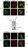
Figure 2. Epidermal growth factor receptor activation induces the co-localization of SH3PXD2B with cortactin and F-actin in membrane ruffles and lamellipodia.
Serum starved cells were left untreated (A–C and H–J) or treated with 100 ng/ml EGF (D–F & K–M) for 10 minutes. Cells were then fixed and processed for immunofluorescence. Endogenous expression of SH3PXD2B and cortactin in HeLa cells (A–F) were detected using SH3PXD2B-specific rabbit polyclonal or cortactin-specific mouse monoclonal antibodies. Cellular localization of SH3PXD2B in Cos7 cells transfected with a construct encoding an SH3PXD2B fusion protein carrying a C-terminal V5 epitope tag (H–M) was visualized using a mouse anti-V5 monoclonal antibody and anti-mouse Alexa488 (green). Filamentous actin (F-actin) was stained with Alexa568-labeled phalloidin. Bars indicate 20 µm. Efficiency of SH3PXD2B-V5 overexpression was analyzed by western blotting (G).
Figure 3
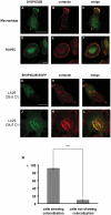
Figure 3. Co-localization of SH3PXD2B with cortactin in HUVEC, macrophages and LA29ts fibroblasts.
Human monocyte-derived macrophages (A–C), HUVEC (D–F) were stained as described for Figure 2A–F. (G–L) Rat-1 LA29 fibroblast cells carrying a temperature sensitive mutant form of Src were grown on coverslips and transfected with an SH3PXD2B-EGFP construct. Cultures were kept at the nonpermissive temperature (39.5°C) for 24 hours. Next, half of the cells were grown further at the nonpermissive temperature until the end of the experiment (G–I). The rest of the cells were cultured at the permissive temperature (34.5°C) overnight (J–L). Coverslips were prepared for immunfuorescence as described in Materials and Methods. Cortactin and SH3PXD2B were visualized as described in the legend for Figure 2. Bars represent 20 µm. Co-localization between SH3PXD2B and cortactin was observed in over 90% of the cells grown at the permissive temperature (M). Over 100 cells were evaluated for each independent experiment. Error bars represent the Standard Error of the Mean, SEM. ***: p<0.001, n = 3.
Figure 4
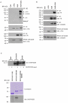
Figure 4. SH3PXD2B associates with Src and cortactin.
(A & B) 293T cells were transfected with constructs expressing full-length SH3PXD2B carrying a C-terminal V5 epitope tag, Src or both. 48 hours past transfection cells were lysed and SH3PXD2B was precipitated using a V5-specific antibody. Immunoprecipitated proteins were blotted onto nitrocellulose membranes and detected with the indicated specific antibodies. The 52 kD-bands present in panel A. correspond to the heavy chain of the antibody used to precipitateV5-tagged SH3PXD2B. (C): In GST pull-down experiments recombinant GST or a GST-cortactin fusion protein were precipitated with glutathion-agarose beads from 293T cell extracts transfected (+) or not (−) with SH3PXD2B (top panel). In similar pull-down experiments GST- fusion proteins representing the N-terminal 334 amino acid region (Cort (1–334)) or the C-terminal region (Cort (336-542)) or its substituent (Cort (336–542(W525K)) were precipitated from lysates of 293T cells (bottom panel). SH3PXD2B was detected by western blotting. GST-proteins (30% of the amounts used in precipitation reactions) are visualized by Coomassie staining (middle panel) Cort: cortactin.
Figure 5
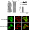
Figure 5. Characterization of the lipid-bindig domain of SH3PXD2B.
(A and B) The lipid specificity of the PX-domain of SH3PXD2B was tested in protein-lipid overlay assay experiments using a recombinant GST fusion protein that contained the first 130 amino acid region of SH3PXD2B (panel A, PX-GST) or its derivative containing the R43Q substitution within the PX-domain (panel B, PX(R43Q). Layout of the PIP strip was the following: 1. Lysophosphatidic acid 2. Lyso-phosphatidylcholine 3. Phosphatidylinositol (PtdIns) 4. PtdIns(3)P 5. PtdIns(4)P 6. PtdIns(5)P 7. Phosphatidylethanolamine 8. Phosphatidylcholine 9. Sphingosine 1-phosphate 10. PtdIns (3,4)P2 11. PtdIns(3,5)P2 12. PtdIns(4,5)P2 13. PtdIns(3,4,5)P3 14. Phosphatidic acid 15. Phosphatidylserine 16. Blank. (C) Liposomes containing Phosphatidylcholine and PtdIns(3)P were mixed with recombinant PX-GST or PX(R43Q)-GST and centrifuged at at 80000 g for 20 minutes. Pellet and supernatant were separated, boiled and then subjected to SDS- polyacrylamide gel electrophoresis and western blotting as described in Materials and Methods. Blots were developed with a GST-specific antibody (top panels). Distribution of the PX-GST proteins between the liposome (pellet) and the aqueous phase (supernatant) was determined by densitometry (bottom panel). (D–I) Constructs expressing wild-type SH3PXD2B-V5 [SH3PXD2B-V5(WT)] or the SH3PXD2B-V5 with the R43Q amino acid substitution [SH3PXD2B-V5(R43Q)] were transfected into COS-7 cells (Panels D–F and G–I, respectively). 24 hours past transfection cells were serum depleted and activated with EGF as described for Figure 2 . Actin and SH3PXD2B-V5 were visualized as described in the legends for Figure 2H–M. Bars represent 20 µm.
Figure 6
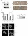
Figure 6. SH3PXD2B is essential for generation of membrane ruffles and lamellipodia.
HeLa cells were grown on coverslips at subconfluent cell densities. Cells were transfected with double stranded control siRNA oligonucleotides (si2c and si3c) or siRNAs specific for SH3PXD2B (si2 and si3). (A) SH3PXD2B levels in cell lysates on the day of the functional assays were determined by western blot analysis. (B–E) At day 2 serum-starved cells were stimulated with EGF, fixed and visualized by phase contrast microscopy. White arrows show membrane ruffles and lamellipodia. (F) EGF-induced ruffling in cells transfected with the indicated siRNA-s were quantified at 10 minutes following activation. Percentages of cells with (columns shaded light grey) or without the characteristic membrane ruffling (columns shaded dark grey) are depicted. 100 cells were evaluated for each independent experiment (n = 4). Error bars represent the Standard Error of the Mean, SEM. ***: p<0.001, **: p<0.005. (G) Expression levels of SH3PXD2B in independent HeLa clones stably expressing SH3PXD2B-specific shRNA (clone 1 and clone 2.) or in two controls (clone 3 and clone 4). EGF-stimulated HeLa cell lines with diminished SH3PXD2B expression (H & I) or with normal SH3PXD2B expression (J & K) were stained for cortactin as described above. White arrows indicate lamellipodia rich in cortactin. Bars represent 20 µm.
Figure 7
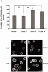
Figure 7. In the absence of SH3PXD2B cell spreading is impaired.
(A–E) The HeLa clones described in Figure 6 were seeded onto fibronectin-coated coverslips, fixed and permeabilized after 30 minutes and stained with TRITC-Phalloidin. Cell surface area was calculated based on images of over 200 cells using the ImageJ 1.38x software. Average size of the cells for two SH3PXD2B-negative (clones 1 and 2) and two control cell lines (clones 3 and 4) is plotted (A). Panels B-E show representative images of poorly spread (B and C) and well spread (D and E) HeLa cells. Error bars represent the Standard Error of the Mean, SEM. ***: p<0.001, n = 6 Bar represents 20 µm.

Similar articles

Cited by

References

    1. Pawson T. Dynamic control of signaling by modular adaptor proteins. Curr Opin Cell Biol. 2007;19:112–116. - PubMed
    1. Pullikuth AK, Catling AD. Scaffold mediated regulation of MAPK signaling and cytoskeletal dynamics: a perspective. Cell Signal. 2007;19:1621–1632. - PMC - PubMed
    1. Ushio-Fukai M. Localizing NADPH oxidase-derived ROS. Sci STKE. 2006. re8 2006. - PubMed
    1. Balla T. Inositol-lipid binding motifs: signal integrators through protein-lipid and protein-protein interactions. J Cell Sci. 2005;118:2093–2104. - PubMed
    1. Lock P, Abram CL, Gibson T, Courtneidge SA. A new method for isolating tyrosine kinase substrates used to identify fish, an SH3 and PX domain-containing protein, and Src substrate. Embo J. 1998;17:4346–4357. - PMC - PubMed

Publication types

MeSH terms