. 2011;6(8):e23703.
doi: 10.1371/journal.pone.0023703. Epub 2011 Aug 23.

Transduction of SIV-specific TCR genes into rhesus macaque CD8+ T cells conveys the ability to suppress SIV replication

Affiliations

Transduction of SIV-specific TCR genes into rhesus macaque CD8+ T cells conveys the ability to suppress SIV replication

Eugene V Barsov et al. PLoS One. 2011.

Erratum in

Abstract

Background: The SIV/rhesus macaque model for HIV/AIDS is a powerful system for examining the contribution of T cells in the control of AIDS viruses. To better our understanding of CD8(+) T-cell control of SIV replication in CD4(+) T cells, we asked whether TCRs isolated from rhesus macaque CD8(+) T-cell clones that exhibited varying abilities to suppress SIV replication could convey their suppressive properties to CD8(+) T cells obtained from an uninfected/unvaccinated animal.

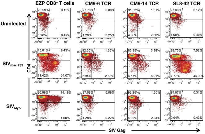
Principal findings: We transferred SIV-specific TCR genes isolated from rhesus macaque CD8(+) T-cell clones with varying abilities to suppress SIV replication in vitro into CD8(+) T cells obtained from an uninfected animal by retroviral transduction. After sorting and expansion, transduced CD8(+) T-cell lines were obtained that specifically bound their cognate SIV tetramer. These cell lines displayed appropriate effector function and specificity, expressing intracellular IFNγ upon peptide stimulation. Importantly, the SIV suppression properties of the transduced cell lines mirrored those of the original TCR donor clones: cell lines expressing TCRs transferred from highly suppressive clones effectively reduced wild-type SIV replication, while expression of a non-suppressing TCR failed to reduce the spread of virus. However, all TCRs were able to suppress the replication of an SIV mutant that did not downregulate MHC-I, recapitulating the properties of their donor clones.

Conclusions: Our results show that antigen-specific SIV suppression can be transferred between allogenic T cells simply by TCR gene transfer. This advance provides a platform for examining the contributions of TCRs versus the intrinsic effector characteristics of T-cell clones in virus suppression. Additionally, this approach can be applied to develop non-human primate models to evaluate adoptive T-cell transfer therapy for AIDS and other diseases.


Conflict of interest statement

Competing Interests: The authors have declared that no competing interests exist.

Figures

Figure 1
Figure 1. Diagram of TCR expressing retroviral vector and the mature TCR chains.
The MSGV1 murine retroviral vectors sequences are displayed as black boxes and lines while the TCR expression cassette is in white. The fine structure of the TCR chain fusion cassette is presented below the vector with the different MamuA*01-restricted TCRs molecularly-cloned from DAJ T-cell clones that were inserted into the vectors indicated above the cassette. The sequences separating the TCR genes, the furin recognition sequence, KAKR, the S-G-S-G spacer, and the P2A fowl pox self-cleaving peptide, are shaded gray. The furin cleavage site and the P2A self-cleavage site are indicated below the cassette with arrows. The mature α and β chains produced by this vector are displayed at the bottom of the figure.
Figure 2
Figure 2. Flow cytometry analysis of transduced T cells.
A, analysis of the TCR-transduced EZP cell lines for CM9 peptide/MHC tetramer and SL8 peptide/MHC tetramer is presented with that of the untransduced CD8+ control cell line from recipient animal EZP. B, tetramer analysis of two SIV-specific CTL clones isolated from donor animal DAJ is presented above tetramer-sorted TCR transduced CD8+ cell lines. The DAJ SL8–42 clone is the TCR gene donor for the SL8–42 TCR EZP cell line.
Figure 3
Figure 3. Intracellular IFNγ assay of TCR-transduced cell lines.
Flow cytometry analysis of intracellular IFNγ production induced by the antigenic peptide is presented. Panel A, an assay of the CM9–6 TCR cell line stimulated with the CM9 peptide is displayed with its corresponding untransduced EZP CD8+ T-cell line control. Panel B, an assay of the CM9–14 TCR and SL8–42 TCR cell lines stimulated with either the CM9 or SL8 peptide is presented below their corresponding untransduced CD8+ T-cell controls. The stimulating peptide used is indicated above the respective plots.
Figure 4
Figure 4. In vitro virus suppression assay of TCR-transduced cell lines.
Flow cytometry analyses of mixed cultures consisting of effector CD8+ T-cell lines and a target autologous CD4+ T-cell clone that was untreated or exposed to either wild-type SIVmac239 or SIVmyr- are presented. Effectors are labeled above each column and targets are labeled at the left of each row. The effector CD8+ T cells in the co-cultures were stained with CellTrace Violet® and excluded from the analysis so that only the target cells were counted.
Figure 5
Figure 5. Suppression of viral RNA load.
A graph of the viral load in the TCR-transduced T-cell mixed cultures relative to those containing untransduced CD8+ T cells is presented. The amount of viral suppression for each culture (viral load of the untransduced CD8+ cells co-culture divided by the load of anti-SIV effector cultures) is indicated above each bar. The average viral loads from duplicate independent PCR-based measurements of the untransduced CD8+ cell co-cultures were 6.6×10⁸ copies of SIV Gag per ml, SD 8.7×10⁶ for wild-type SIVmac239 and 3.8×10⁸ copies of SIV Gag per ml, SD 1.7×10⁷ for the myristylation mutant SIVmyr-. Error bars represent standard deviations.

Similar articles

Cited by

References

    1. Bollard CM, Aguilar L, Straathof KC, Gahn B, Huls MH, et al. Cytotoxic T lymphocyte therapy for Epstein-Barr virus+ Hodgkin's disease. J Exp Med. 2004;200:1623–1633. - PMC - PubMed
    1. Dudley ME, Wunderlich JR, Robbins PF, Yang JC, Hwu P, et al. Cancer regression and autoimmunity in patients after clonal repopulation with antitumor lymphocytes. Science. 2002;298:850–854. - PMC - PubMed
    1. Dudley ME, Wunderlich JR, Yang JC, Sherry RM, Topalian SL, et al. Adoptive cell transfer therapy following non-myeloablative but lymphodepleting chemotherapy for the treatment of patients with refractory metastatic melanoma. J Clin Oncol. 2005;23:2346–2357. - PMC - PubMed
    1. Rooney CM, Smith CA, Ng CY, Loftin SK, Sixbey JW, et al. Infusion of cytotoxic T cells for the prevention and treatment of Epstein-Barr virus-induced lymphoma in allogeneic transplant recipients. Blood. 1998;92:1549–1555. - PubMed
    1. Walter EA, Greenberg PD, Gilbert MJ, Finch RJ, Watanabe KS, et al. Reconstitution of cellular immunity against cytomegalovirus in recipients of allogeneic bone marrow by transfer of T-cell clones from the donor. N Engl J Med. 1995;333:1038–1044. - PubMed

Publication types

MeSH terms

Substances