. 2011;6(8):e23706.
doi: 10.1371/journal.pone.0023706. Epub 2011 Aug 19.

The targeting of plasmalemmal ceramide to mitochondria during apoptosis

Affiliations

The targeting of plasmalemmal ceramide to mitochondria during apoptosis

Eduard B Babiychuk et al. PLoS One. 2011.

Abstract

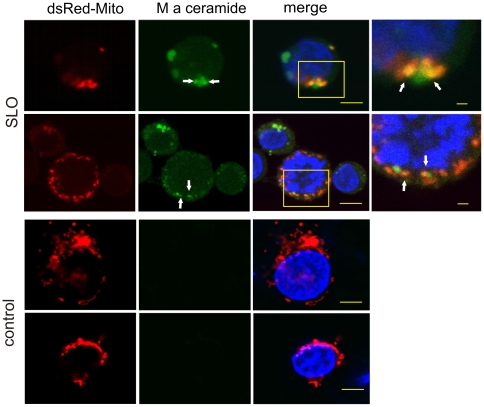
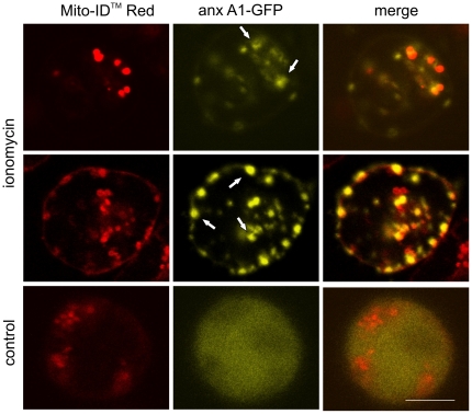
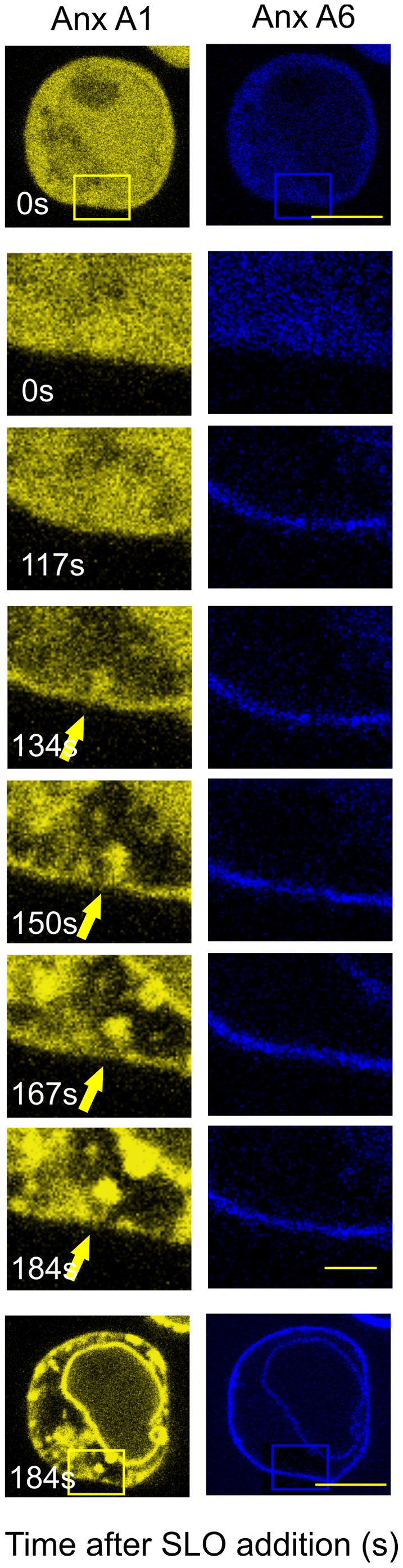
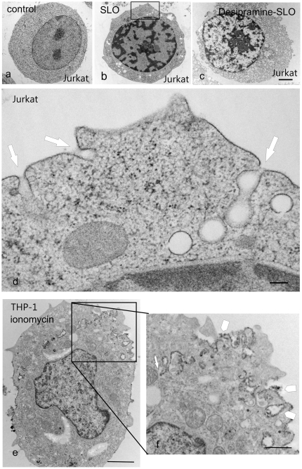
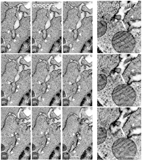
Ceramide is a key lipid mediator of cellular processes such as differentiation, proliferation, growth arrest and apoptosis. During apoptosis, ceramide is produced within the plasma membrane. Although recent data suggest that the generation of intracellular ceramide increases mitochondrial permeability, the source of mitochondrial ceramide remains unknown. Here, we determine whether a stress-mediated plasmalemmal pool of ceramide might become available to the mitochondria of apoptotic cells. We have previously established annexin A1--a member of a family of Ca(2+) and membrane-binding proteins--to be a marker of ceramide platforms. Using fluorescently tagged annexin A1, we show that, upon its generation within the plasma membrane, ceramide self-associates into platforms that subsequently invaginate and fuse with mitochondria. An accumulation of ceramide within the mitochondria of apoptotic cells was also confirmed using a ceramide-specific antibody. Electron microscopic tomography confirmed that upon the formation of ceramide platforms, the invaginated regions of the plasma membrane extend deep into the cytoplasm forming direct physical contacts with mitochondrial outer membranes. Ceramide might thus be directly transferred from the plasma membrane to the mitochondrial outer membrane. It is conceivable that this "kiss-of-death" increases the permeability of the mitochondrial outer membrane thereby triggering apoptosis.


Conflict of interest statement

Competing Interests: The authors have declared that no competing interests exist.

Figures

Figure 1
Figure 1. Ca2+-overload leads to the appearance of ceramide in mitochondria.
The mitochondrial marker dsRed-Mito (red) was transiently expressed in Jurkat T-cells, which were either stimulated with SLO or left untreated (control). Subsequently, the cells were chemically fixed and treated with a polyclonal antibody against ceramide (M a ceramide; green) and Hoechst stain as a nuclear marker (blue). Confocal micrographs revealed ceramide platforms and their partial colocalization with mitochondria in stimulated cells (yellow, arrows). The boxed areas are enlarged. No ceramide platforms are present in unstimulated cells. Bars in the merged images = 3 µm, enlarged boxed areas = 1 µm.
Figure 2
Figure 2. Annexin A1 partially colocalises with mitochondria after intracellular Ca2+-overload.
Confocal micrographs of living Jurkat T-cells that had been transiently transfected with annexin A1-GFP, treated with the mitochondrial marker Mito-ID™Red (red) and stimulated with ionomycin. A partial co-localization of annexin A1 with mitochondria can be observed (arrows). The untreated cell (control) shows a homogeneous distribution of annexin A1 throughout the cytoplasm. Bar = 3 µm.
Figure 3
Figure 3. Annexin A1 invagination in a Jurkat T-cell after intracellular Ca2+-overload.
Time-lapse sequence of confocal micrographs for a Jurkat T-cell that had been transiently transfected with annexin A1-YFP (Anx A1, yellow) and annexin A6 (Anx A6, blue) prior to stimulation with SLO. Both annexins translocate to the plasma membrane between time points 117–134. Thereafter, annexin A1 segregates from annexin A6 and coalesces into membrane platforms, which are internalised. The development of annexin A1-decorated, finger-like invaginations after Ca2+overload (arrows) was monitored over 3 min (time in s). Images at selected time-points are illustrated. Those at time-points zero and 184s (bars = 3 µm) are represented at lower magnification (boxed areas; bar = 1 µm) to aid orientation.
Figure 4
Figure 4. Tube-like invagination of the plasma membrane after intracellular Ca2+-overload.
Electron micrographs of unfixed, high pressure frozen, freeze-substituted Jurkat T cells and THP-1 cells at rest (a), after stimulation with SLO (b,d) and after treatment with sphingomyelinase inhibitor desipramine before stimulation with SLO (c). (e,f ) THP-1 cells were pre-labelled with horseradish peroxidase-cholera toxin B (arrowheads) before treatment with ionomycin. a: The untreated (control) cell displays a homogeneous cytoplasm and a uniformally smooth plasmalemmal surface. b,d: The SLO-stimulated cell contains numerous cytoplasmic vacuolations, which at higher resolution [d (boxed area in b] turn out to be the profiles of tubular invaginations of the plasma membrane (arrows). c: For the inhibition of sphingomyelinase, the cells were preincubated at 37°C for 90 minutes in the presence of 50 µM desipramine prior to stimulation with SLO. e: please note deep surface-labelled plasma membrane invaginations, boxed area enlarged in (f, arrow) Bars: a,b,c,e = 1 µm, d = 0.2 µm, f = 0.5 µm.
Figure 5
Figure 5. “Kiss-of-death” between the plasma membrane and mitochondria.
Movie stills of electron micrographic tomograms of 2 different, unfixed, frozen Jurkat cells, whose outer leaflets of the plasma membrane were pre-labelled with horseradish peroxidase-cholera toxin B (arrowheads) before subjecting the cells to Ca2+ overload. Contact sites between surface-labelled membrane invaginations which extend deeply into the cytoplasm and mitochondrial outer membranes are visible (arrows). Mitochondrium (m), Nucleus (N). Bars = 0.5 µm.

References

    1. Bleicken S, Classen M, Padmavathi PV, Ishikawa T, Zeth K, et al. Molecular details of Bax activation, oligomerization, and membrane insertion. J Biol Chem. 2010;285:6636–6647. - PMC - PubMed
    1. Green DR, Kroemer G. The pathophysiology of mitochondrial cell death. Science. 2004;305:626–629. - PubMed
    1. George NM, Targy N, Evans JJ, Zhang L, Luo X. Bax contains two functional mitochondrial targeting sequences and translocates to mitochondria in a conformational change- and homo-oligomerization-driven process. J Biol Chem. 2010;285:1384–1392. - PMC - PubMed
    1. Martinez-Abundis E, Correa F, Pavon N, Zazueta C. Bax distribution into mitochondrial detergent-resistant microdomains is related to ceramide and cholesterol content in postischemic hearts. FEBS J. 2009;276:5579–5588. - PubMed
    1. Tait SW, Green DR. Mitochondria and cell death: outer membrane permeabilization and beyond. Nat Rev Mol Cell Biol. 2010;11:621–632. - PubMed

Publication types