

Air trapping on chest CT is associated with worse ventilation distribution in infants with cystic fibrosis diagnosed following newborn screening
- PMID: 21886842
- PMCID: PMC3158781
- DOI: 10.1371/journal.pone.0023932
Air trapping on chest CT is associated with worse ventilation distribution in infants with cystic fibrosis diagnosed following newborn screening
Abstract
Background: In school-aged children with cystic fibrosis (CF) structural lung damage assessed using chest CT is associated with abnormal ventilation distribution. The primary objective of this analysis was to determine the relationships between ventilation distribution outcomes and the presence and extent of structural damage as assessed by chest CT in infants and young children with CF.
Methods: Data of infants and young children with CF diagnosed following newborn screening consecutively reviewed between August 2005 and December 2009 were analysed. Ventilation distribution (lung clearance index and the first and second moment ratios [LCI, M(1)/M(0) and M(2)/M(0), respectively]), chest CT and airway pathology from bronchoalveolar lavage were determined at diagnosis and then annually. The chest CT scans were evaluated for the presence or absence of bronchiectasis and air trapping.
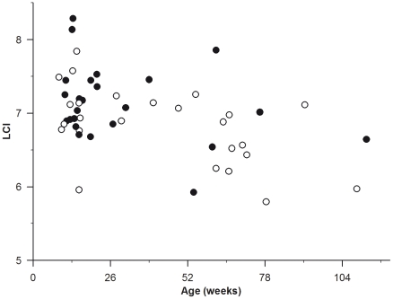
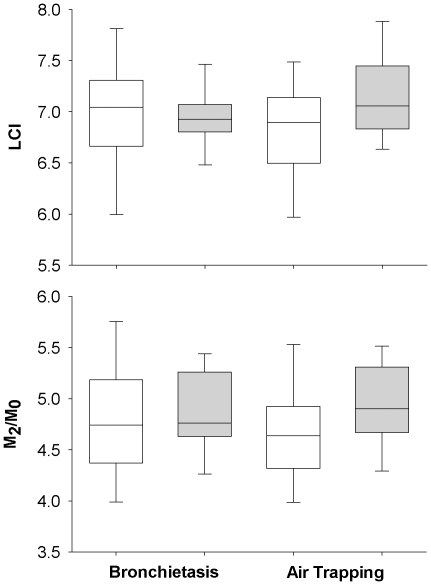
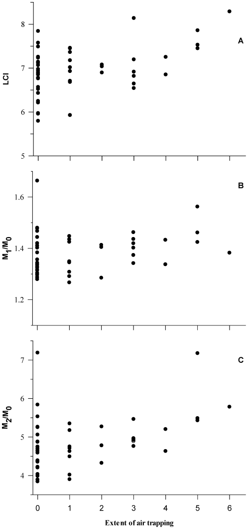
Results: Matched lung function, chest CT and pathology outcomes were available in 49 infants (31 male) with bronchiectasis and air trapping present in 13 (27%) and 24 (49%) infants, respectively. The presence of bronchiectasis or air trapping was associated with increased M(2)/M(0) but not LCI or M(1)/M(0). There was a weak, but statistically significant association between the extent of air trapping and all ventilation distribution outcomes.
Conclusion: These findings suggest that in early CF lung disease there are weak associations between ventilation distribution and lung damage from chest CT. These finding are in contrast to those reported in older children. These findings suggest that assessments of LCI could not be used to replace a chest CT scan for the assessment of structural lung disease in the first two years of life. Further research in which both MBW and chest CT outcomes are obtained is required to assess the role of ventilation distribution in tracking the progression of lung damage in infants with CF.
Conflict of interest statement
Figures
References
-
- Linnane B, Robinson P, Ranganathan S, Stick S, Murray C. Role of high-resolution computed tomography in the detection of early cystic fibrosis lung disease. Paediatr Respir Rev. 2008;9:168–174; quiz 174–165. - PubMed
-
- Tiddens HA. Chest computed tomography scans should be considered as a routine investigation in cystic fibrosis. Paediatr Respir Rev. 2006;7:202–208. - PubMed
-
- Davis SD, Fordham LA, Brody AS, Noah TL, Retsch-Bogart GZ, et al. Computed tomography reflects lower airway inflammation and tracks changes in early cystic fibrosis. Am J Respir Crit Care Med. 2007;175:943–950. - PubMed
-
- Stick SM, Brennan S, Murray C, Douglas T, von Ungern-Sternberg BS, et al. Bronchiectasis in infants and preschool children diagnosed with cystic fibrosis after newborn screening. J Pediatr. 2009;155:623–628. - PubMed
Publication types
MeSH terms
LinkOut - more resources
Full Text Sources
Medical
