. 2011:2:107.
doi: 10.4103/2152-7806.83391. Epub 2011 Jul 30.

Immunoexcitotoxicity as a central mechanism in chronic traumatic encephalopathy-A unifying hypothesis

Affiliations

Immunoexcitotoxicity as a central mechanism in chronic traumatic encephalopathy-A unifying hypothesis

Russell L Blaylock et al. Surg Neurol Int. 2011.

Abstract

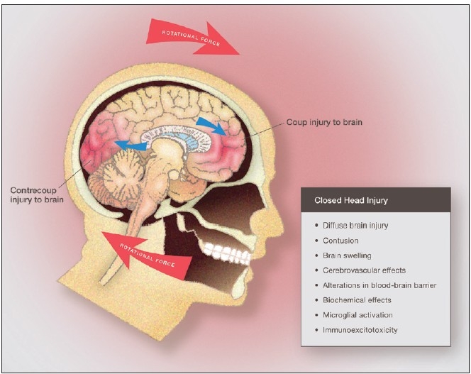
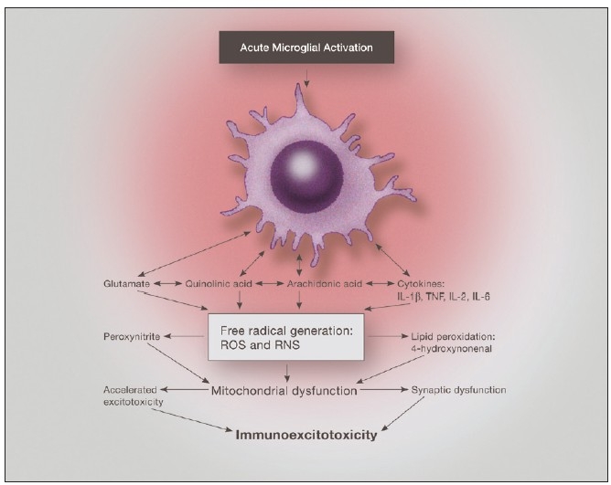
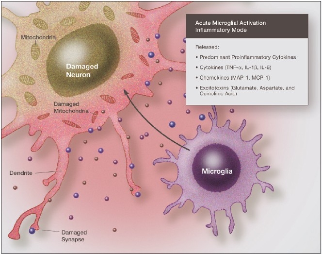
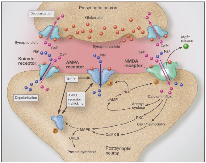
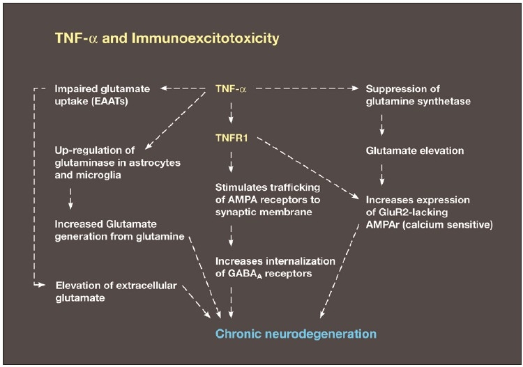
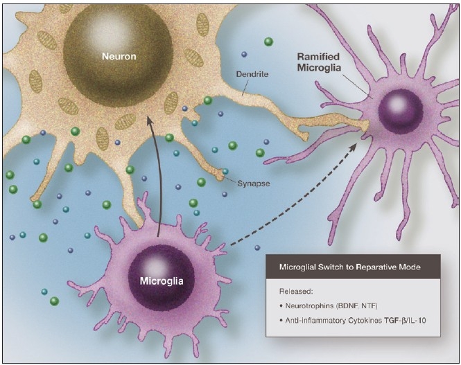
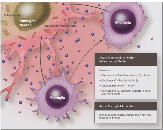
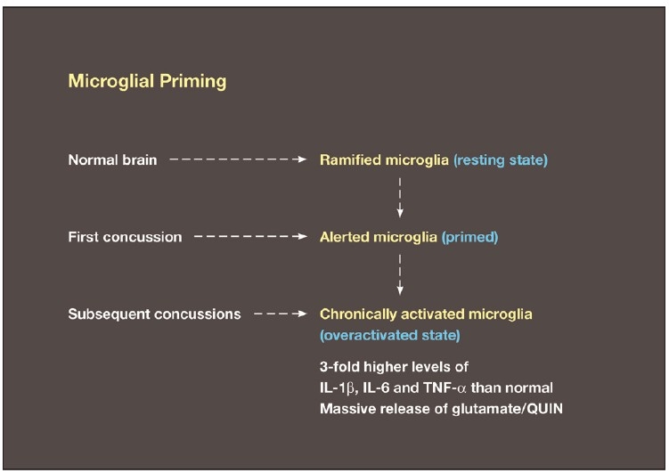
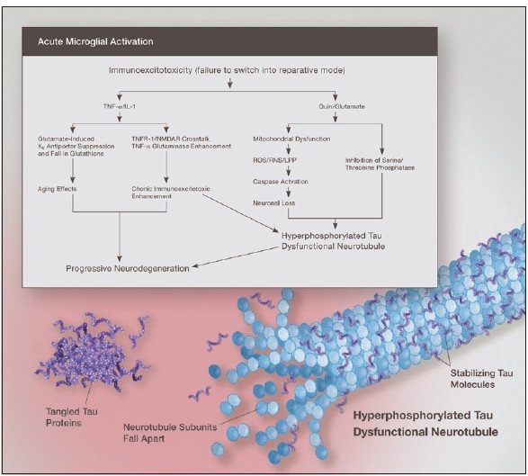
Some individuals suffering from mild traumatic brain injuries, especially repetitive mild concussions, are thought to develop a slowly progressive encephalopathy characterized by a number of the neuropathological elements shared with various neurodegenerative diseases. A central pathological mechanism explaining the development of progressive neurodegeneration in this subset of individuals has not been elucidated. Yet, a large number of studies indicate that a process called immunoexcitotoxicity may be playing a central role in many neurodegenerative diseases including chronic traumatic encephalopathy (CTE). The term immunoexcitotoxicity was first coined by the lead author to explain the evolving pathological and neurodevelopmental changes in autism and the Gulf War Syndrome, but it can be applied to a number of neurodegenerative disorders. The interaction between immune receptors within the central nervous system (CNS) and excitatory glutamate receptors trigger a series of events, such as extensive reactive oxygen species/reactive nitrogen species generation, accumulation of lipid peroxidation products, and prostaglandin activation, which then leads to dendritic retraction, synaptic injury, damage to microtubules, and mitochondrial suppression. In this paper, we discuss the mechanism of immunoexcitotoxicity and its link to each of the pathophysiological and neurochemical events previously described with CTE, with special emphasis on the observed accumulation of hyperphosphorylated tau.

Keywords: Cerebral concussion; chronic traumatic encephalopathy; cytokines; hyperphosphorylated tau; immunoexcitotoxicity; microglia; mild traumatic brain injury; quniolinic acid.


Figures

Figure 1
Figure 1
Illustration of closed brain injury demonstrating mechanical forces involved and the pathophysiological and biochemical effects of diffuse brain injury, which result in rapid microglial activation and immunoexcitotoxicity
Figure 2
Figure 2
Illustration of the neurotoxic factors released from an activated microglia, demonstrating the interaction of proinflammatory cytokines and excitatory amino acids. Of particular importance is the effect on mitochondrial function, which when depressed enhances excitotoxic sensitivity as well as reactive oxygen species generation
Figure 3
Figure 3
Illustration demonstrating the neurotoxic effects of proinflammatory cytokines and chemokines acting in synergy with excitatory amino acids. Immunoexcitoxicity results in damage to neuronal cell membranes, mitochondria, DNA, as well as dendrites and synapses
Figure 4
Figure 4
Illustration of glutamatergic synapse demonstrating AMPA receptor trafficking from the endoplasmic reticulum, which is driven by activation of tumor necrosis factor receptor-1. Crosstalk between the 2-amino-3-(5-methyl-3-oxo-1,2-oxazol-4-yl) propanoic acid receptor and tumor necrosis factor receptor-1 increase synaptic insertion of GluR2-lacking (calcium permeable) 2-amino-3-(5-methyl-3-oxo-1,2-oxazol-4-yl) propanoic acid receptors, thus increasing synaptic glutamate-related sensitivity. tumor necrosis factor receptor-1 activation also increases GABA receptor endocytosis, which increases synaptic sensitivity to excitotoxicity even further
Figure 5
Figure 5
Diagram demonstrating a number of the major mechanisms of immunoexcitotoxicity, which includes the interaction of TNF-α with a number of systems that enhance excitotoxicity. This includes impaired glutamate transport, upregulation of glutaminase, suppression of glutamine synthetase, increased trafficking of AMPA receptors to synaptic lipid raft and endocytosis of GABA
Figure 6
Figure 6
Illustration of microglia priming/activation transition states beginning from a resting (ramified) state. Recent studies indicate that microglia can assume a number of activation states, such as predominately phagocytic, predominately neuroprotective or predominately neurodestructive. In the primed state the mRNA for cytokines, chemokines and other reactive molecules are upregulated but active proteins are not released
Figure 7
Figure 7
Illustration of a microglia in a predominately reparative mode, which will then switch to a resting (ramified) state. In the reparative mode it secretes neurotrophic factors and anti-inflammatory cytokines that shut off the inflammatory reaction
Figure 8
Figure 8
Illustration of an activated microglia that fails to switch from an activated, neurodestructive mode to a reparative mode or ramified state. Under such conditions immunoexcitotoxic reactions can continue for prolonged periods
Figure 9
Figure 9
Diagram demonstrating the conversion of a resting microglia in the uninjured brain to a primed microglia with an initial injury. Subsequent injuries, even separated by prolong periods, can then trigger a hyper-reaction by the fully activated microglia. This in turn results in a more intense immunoexcitotoxic reaction
Figure 10
Figure 10
Elevated glutamate and subsequent excitotoxicity is essential to neurodegeneration induced by elevated proinflammatory cytokines. With chronic brain inflammation, tryptophan metabolism by the kynurenine pathway shifts toward quinolinic acid generation, which is excitotoxic. Both elevated QUIN and glutamate levels inhibit phosphatases and this results in hyperphosphorylated tau and subsequent neurotubule dysfunction and neurofibrillary tangle deposition in the areas of the brain most affected by immunoexcitotoxicity

References

    1. Adams JH, Doyle D, Ford I, Gennarelli TA, Graham DI, McClellan DR. Diffuse axonal injury in head injury: definition, diagnosis and grading. Histopathology. 1989;15:49–59. - PubMed
    1. Adams JH, Graham DI, Gennarelli TA, Maxwell WL. Diffuse axonal injury in non-missile head injury. J Neurol Neurosurg Psychaiatry. 1991;54:481–3. - PMC - PubMed
    1. Alnoni S, Cervia D, Ross B, Montanari C, Gonzales AS, Sanchez-Alvez M, et al. Mapping of the full length and the truncated interleukin-18 receptor alpha in the mouse brain. J Neuroimmunol. 2009;214:43–54. - PMC - PubMed
    1. Andersson PB, Perry VH, Gordon S. Intracerebral injection of proinflammatory cytokines or leukocyte chemotaxins induces minimal myelomonocytic cell recruitment to the parenchyma of the central nervous system. Brain. 1997;120:435–44. - PMC - PubMed
    1. Anisman H. Cascading effects of stressors and inflammatory immune system activation: implication for major depressive disorder. Rev Psychiatry Neurosci. 2009;34:4–20. - PMC - PubMed