

Validation of a flow cytometry based binding assay for evaluation of monoclonal antibody recognizing EGF receptor
- PMID: 21886904
- PMCID: PMC3163362
- DOI: 10.3797/scipharm.1104-18
Validation of a flow cytometry based binding assay for evaluation of monoclonal antibody recognizing EGF receptor
Abstract
An ideal test used to characterize a product must be appropriate for the measurement of product quality, manufacturing consistency, product stability, and comparability studies. Flow cytometry has been successfully applied to the examination of antibodies and receptors on membrane surfaces; however, to date, the analytical validation of cytometry based assays is limited. Here we report on the validation of a flow cytometry-based assay used in the evaluation of nimotuzumab binding to cells over-expressing EGFR on cell surface. The assay was validated by examining, assay robustness, specificity, repeatability and intermediate precision. The assay was highly specific, robust for all studied factors except for cell fixation with 1% paraformaldehyde and met criteria for precision with RSD < 2%. In addition the assay has stability-indicating properties evidenced by the ability to detect changes in mAb degraded samples. Most importantly, the assay demonstrated to be useful for its intended use.
Keywords: Binding Assays; Method validation; Monoclonal antibody; Nimotuzumab.
Figures
 ), degraded nimotuzumab (
 ), native nimotuzumab (
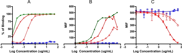
 ), or native FITC-conjugated nimotuzumab (
 ), antihuman/FITC was used as secondary Ab when non-conjugated mAb was evaluated. [C], Results of competition binding assay, concentrations from 0.01 to 50 μg/mL of native and degraded mAbs were incubated for 30 min at 4°C in the presence of FITC-Conjugate nimotuzumab (2.5 μg/mL).
Similar articles
-
Validation of a flow cytometry based chemokine internalization assay for use in evaluating the pharmacodynamic response to a receptor antagonist.J Transl Med. 2008 Dec 1;6:76. doi: 10.1186/1479-5876-6-76. J Transl Med. 2008. PMID: 19046455 Free PMC article. Clinical Trial.
-
Validation of a HLA-A2 tetramer flow cytometric method, IFNgamma real time RT-PCR, and IFNgamma ELISPOT for detection of immunologic response to gp100 and MelanA/MART-1 in melanoma patients.J Transl Med. 2008 Oct 22;6:61. doi: 10.1186/1479-5876-6-61. J Transl Med. 2008. PMID: 18945350 Free PMC article.
-
Validation of a flow cytometry based G(2)M delay cell cycle assay for use in evaluating the pharmacodynamic response to Aurora A inhibition.J Immunol Methods. 2011 Jan 5;363(2):135-42. doi: 10.1016/j.jim.2010.09.021. Epub 2010 Sep 29. J Immunol Methods. 2011. PMID: 20887727
-
Development of ABX-EGF, a fully human anti-EGF receptor monoclonal antibody, for cancer therapy.Crit Rev Oncol Hematol. 2001 Apr;38(1):17-23. doi: 10.1016/s1040-8428(00)00134-7. Crit Rev Oncol Hematol. 2001. PMID: 11255078 Review.
-
Validation of serological assays for diagnosis of infectious diseases.Rev Sci Tech. 1998 Aug;17(2):469-526. doi: 10.20506/rst.17.2.1119. Rev Sci Tech. 1998. PMID: 9713892 Review. English, French, Spanish.
Cited by
-
Receptor occupancy assessment by flow cytometry as a pharmacodynamic biomarker in biopharmaceutical development.Cytometry B Clin Cytom. 2016 Mar;90(2):117-27. doi: 10.1002/cyto.b.21259. Epub 2015 Jul 31. Cytometry B Clin Cytom. 2016. PMID: 26054054 Free PMC article. Review.
-
Polymalic acid chlorotoxin nanoconjugate for near-infrared fluorescence guided resection of glioblastoma multiforme.Biomaterials. 2019 Jun;206:146-159. doi: 10.1016/j.biomaterials.2019.03.029. Epub 2019 Mar 23. Biomaterials. 2019. PMID: 30933776 Free PMC article.
-
Applications of Surface Plasmon Resonance and Biolayer Interferometry for Virus-Ligand Binding.Viruses. 2022 Mar 29;14(4):717. doi: 10.3390/v14040717. Viruses. 2022. PMID: 35458446 Free PMC article. Review.
References
-
- International conference on the harmonization (ICH) Specifications: test procedures and acceptance criteria for biotechnological/biological products. Q6B. 1999.
-
- International conference on the harmonization (ICH) Validation of analytical procedures, Q2 (R1): Text and Methodology. Step 4, 2005.
-
- Food and Drug Administration (FDA) Guidance for Industry. Bioanalytical Method Validation. 2001. pp. 2–4.
LinkOut - more resources
Full Text Sources
Other Literature Sources
Research Materials
Miscellaneous